

How Data Powers AI Innovation in Radiology: Insights & Challenges
Modern artificial intelligence (AI) solutions offered by the main vendors on the market are fundamentally built upon machine learning (ML) and deep learning (DL) algorithms that are inherently data-driven. The success of an AI applications in radiology hinges on the availability of extensive libraries; data are crucial for training, validating, and continually enhancing AI models.
Introduction
In the realm of radiology—where data encompass a complex array of medical images, textual information, and various forms of structured and unstructured data—the quality, diversity, and quantity of data directly affect the effectiveness of AI algorithms. This article touches on the pivotal role that data play in AI solutions for radiology. We will explain the complexities of data storage, management, security, and privacy, while also highlighting the ethical considerations involved in data utilization within AI, for you to better understand how different vendors confront this sensible subject.
Types and Sources of Data in Radiology
Medical Imaging Data: The primary data in radiology consist of medical images such as X-rays, computed tomography (CT) scans, magnetic resonance imaging (MRI), positron emission tomography (PET) scans, and ultrasound (US) images. These images, often enriched with metadata like patient demographics, medical histories, and imaging parameters, serve as the foundational input for AI algorithms.

Clinical Information offer a rich context for AI algorithms to interpret and analyze medical images effectively. This information includes both structured and unstructured data that enhance the diagnostic capabilities of AI models.
Structured Data refer to highly organized information typically stored in relational databases or spreadsheets. In radiology, this encompasses patient demographics, laboratory results, diagnostic and procedure codes, and summaries from radiology reports. Because they are easily searchable and analyzable, structured data provide valuable supplementary information for image analysis, adding depth to the AI's understanding of medical images.
Unstructured Data in radiology mainly consist of textual content such as radiology reports, clinical notes, and referral letters. Despite lacking formal organization, these data contain a wealth of information that can be harnessed using natural language processing (NLP) techniques. AI algorithms can extract meaningful insights from this unstructured text to support and enhance image interpretation.
Additional Data Sources: Beyond the primary data types, several other sources can further enrich AI solutions in radiology:
- Electronic Health Records (EHRs): These records encompass a vast array of both structured and unstructured clinical data, providing a comprehensive view of a patient's medical history.
- Genomic Data: Information about a patient's genetic makeup can influence disease prognosis and treatment responses, offering personalized insights that can be integrated into AI models.
- Medical Literature: The continually expanding body of medical research can be incorporated into AI algorithms to ensure that the most up-to-date, evidence-based practices inform diagnostic and treatment recommendations.
Data Collection Methods
Traditionally, the radiology department has depended on the expertise of doctors who carefully interpret medical images and annotate findings. This manual annotation process involves labeling images to identify and outline areas of interest or abnormalities and forms the basis of radiology reports, which are essential clinical documents.
Unfortunately, manual data collection and annotation come with limitations. The process is time-consuming, subject to variability between different observers, and can be influenced by subjective judgments. Additionally, the sheer volume of data generated in radiology can be overwhelming, potentially leading to delays and inaccuracies in interpretation.
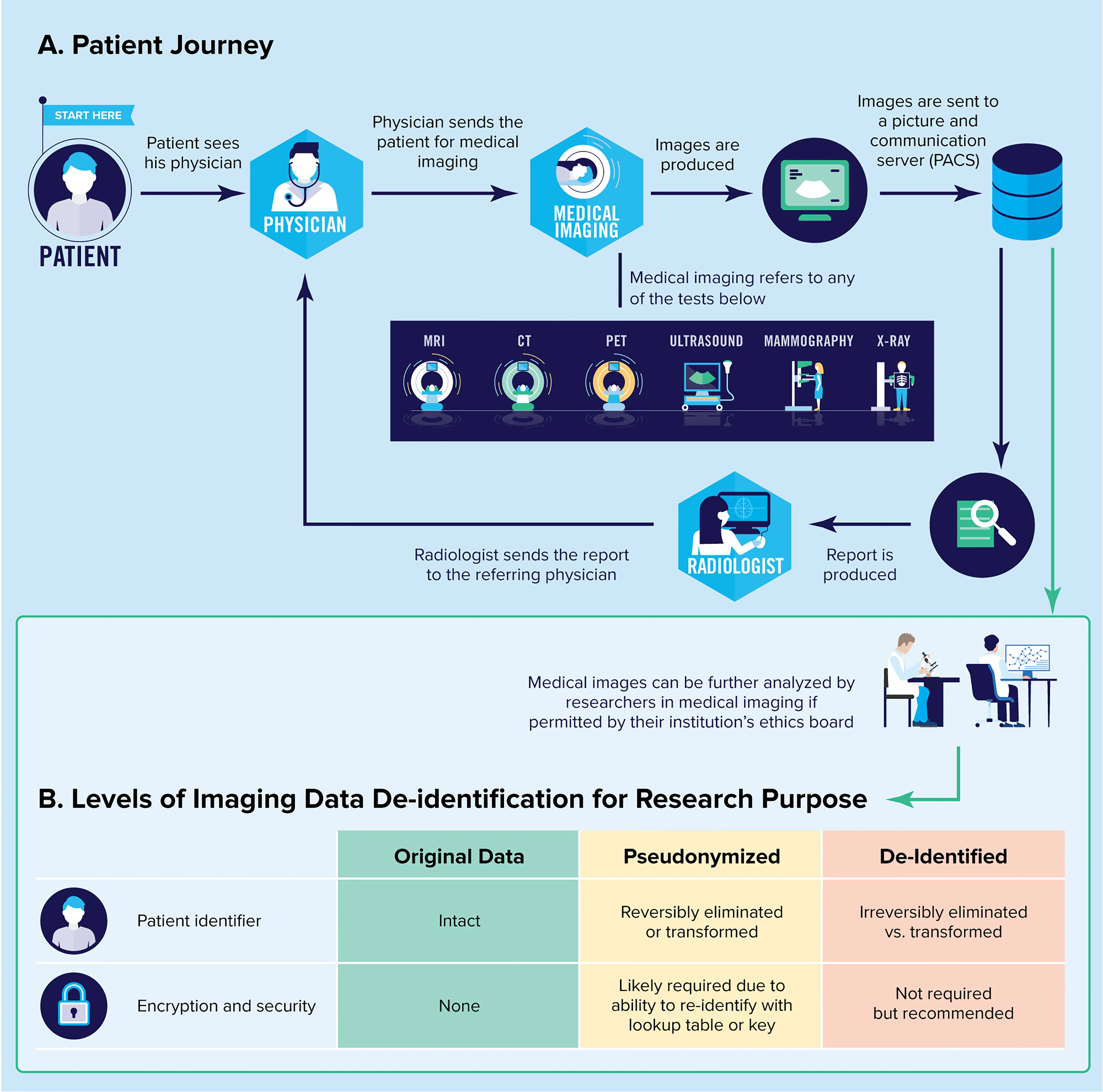
To address these challenges, advancements in data collection techniques have been developed. The implementation of digital imaging systems and the widespread adoption of Picture Archiving and Communication Systems (PACS) have significantly improved efficiency. PACS allow for the digital storage and retrieval of medical images, facilitating easy access and sharing across various clinical settings.

The standardization of medical imaging through the Digital Imaging and Communications in Medicine (DICOM) standards has revolutionized data handling. DICOM provides a uniform framework for storing, transmitting, and printing medical images along with patient information, making it an invaluable asset for AI applications in radiology.
These technological advancements have greatly expanded the potential of AI in radiology by enabling the mass digitization and standardization of medical images. They create large, diverse datasets that are readily accessible for training and refining AI models, thereby enhancing their performance and reliability.
The Role of Data Annotation
Data annotation is a crucial process for an AI vendor as it involves adding labels to data to identify specific features or regions of interest, such as anatomical structures or lesions. This annotated data serves as the "ground truth" from which AI algorithms learn patterns and make predictions. For example, expert radiologists might outline the boundaries of tumors on a series of MRI scans to teach an AI model how to recognize similar features.
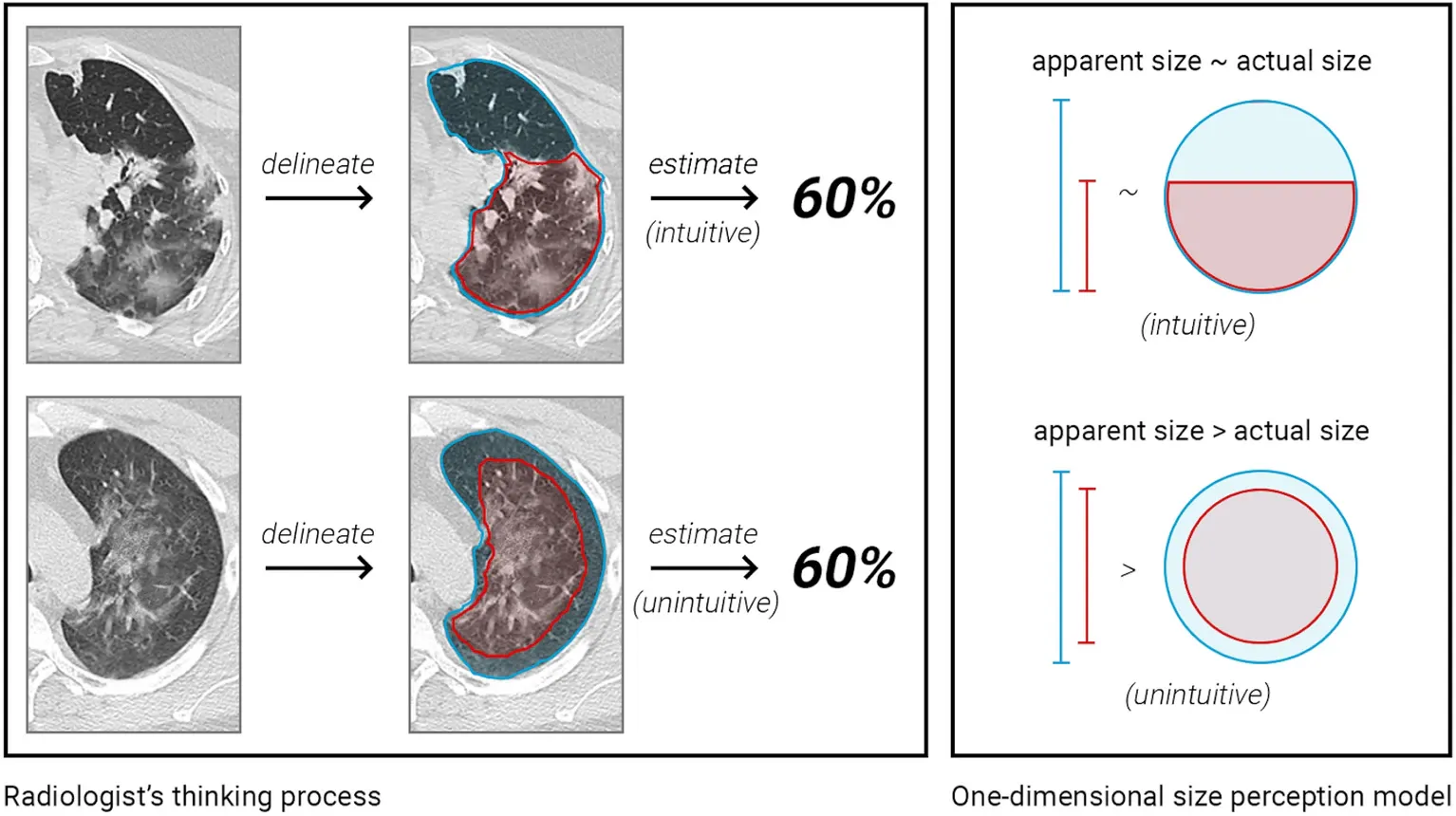
While manual annotation is invaluable for its accuracy and detail, it is also labor-intensive, expensive and subject to differences between annotators. To mitigate these issues, semi-automatic and automatic annotation tools are being developed. These tools aim to streamline the annotation process by using algorithms to suggest annotations, which experts can then review and adjust as needed.
Data annotation is not limited to imaging data; it is equally important for other data types used in AI applications. For structured data, annotation might involve tagging data points with labels that indicate their medical significance, such as marking lab results that signify a particular condition.
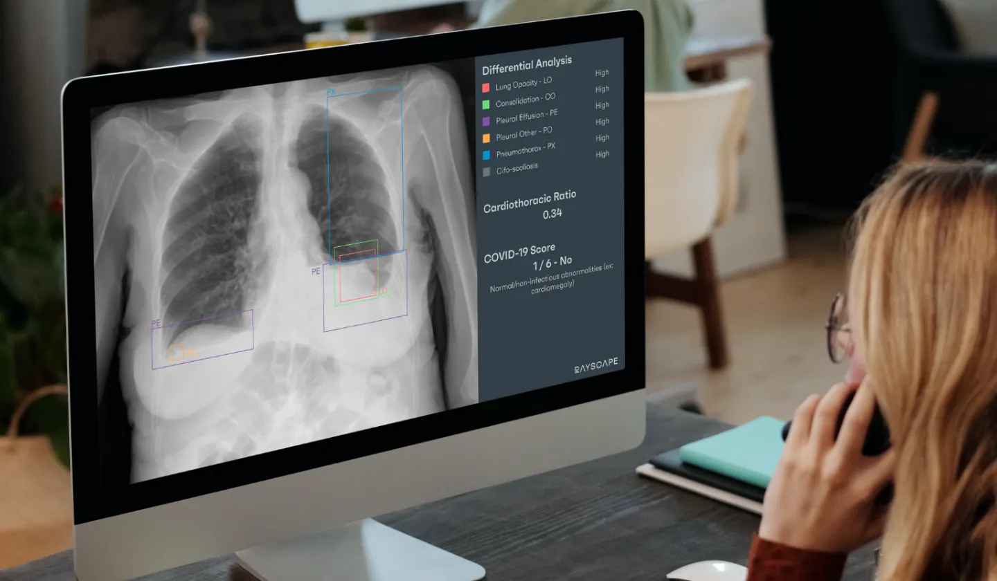
For unstructured text data, annotation often requires NLP techniques to extract and label relevant information, such as identifying medical terms, patient outcomes, or categorizing reports based on findings. This transforms free-text data into structured formats that AI algorithms can effectively utilize.
Data Challenges and Critical Considerations
When developing AI solutions in radiology, several data-related challenges need to be addressed by the vendor to ensure the effectiveness and fairness of these technologies. Key issues include data scarcity, heterogeneity, bias, and concerns surrounding security and privacy.
1) Data Scarcity: One of the most prevalent challenges is the limited availability of labeled and annotated datasets, especially for rare diseases or complex medical conditions. This shortage can hinder the development and training of accurate and robust AI models. Insufficient data may lead to overfitting, where a model performs well on training data but fails to generalize to new, unseen cases.
To overcome data scarcity, several strategies can be employed:
- Data Augmentation: This technique involves expanding the existing dataset by applying various transformations—such as rotations, translations, or scaling—to the images. By increasing the diversity and variability within the dataset, data augmentation enhances the robustness and performance of AI algorithms.
- Synthetic Data Generation: Utilizing methods like generative adversarial networks (GANs), artificial data samples that closely resemble real-world data can be created. These synthetic examples can supplement limited or unbalanced datasets, enabling more effective training of AI models.
- Collaboration and Data Sharing: Pooling data resources through collaboration among healthcare institutions can lead to more extensive and diverse datasets. Privacy-preserving approaches like federated learning allow for data sharing without compromising patient confidentiality, addressing data scarcity while maintaining security.
2) Data Heterogeneity refers to the diversity in data sources, formats, and characteristics, which poses significant challenges in integrating and analyzing medical data. Variations in imaging protocols, equipment types, and patient populations can introduce inconsistencies and biases, affecting the performance and generalizability of AI models.
To address these issues:
- Standardization: Establishing common data formats, ontologies, and metadata standards promotes consistent data representation. This facilitates seamless integration of diverse data sources and enhances compatibility across different systems.
- Data Harmonization: Implementing techniques to map different terminologies and normalize imaging protocols helps mitigate the impact of heterogeneity. By aligning data from various sources to a common standard, it becomes easier to compare and combine datasets effectively.
3) Data Bias: Bias in data arises from systematic errors or favoritism during data collection and annotation. Factors contributing to data bias include demographic disparities, inconsistent data collection practices, and subjective human annotations. Biased data can lead to unfair predictions and inequitable outcomes, undermining the fairness and accuracy of AI solutions.
Strategies to mitigate data bias include:
- Diverse and Representative Datasets: Collecting data that encompass a wide range of demographics, clinical presentations, and imaging characteristics helps minimize bias resulting from underrepresented groups.
- Fairness-Aware Modeling Techniques: Incorporating methods during model development that assess and correct for bias can promote equitable decision-making. These techniques involve evaluating model predictions for potential biases and implementing adjustments to ensure fairness across different patient populations.
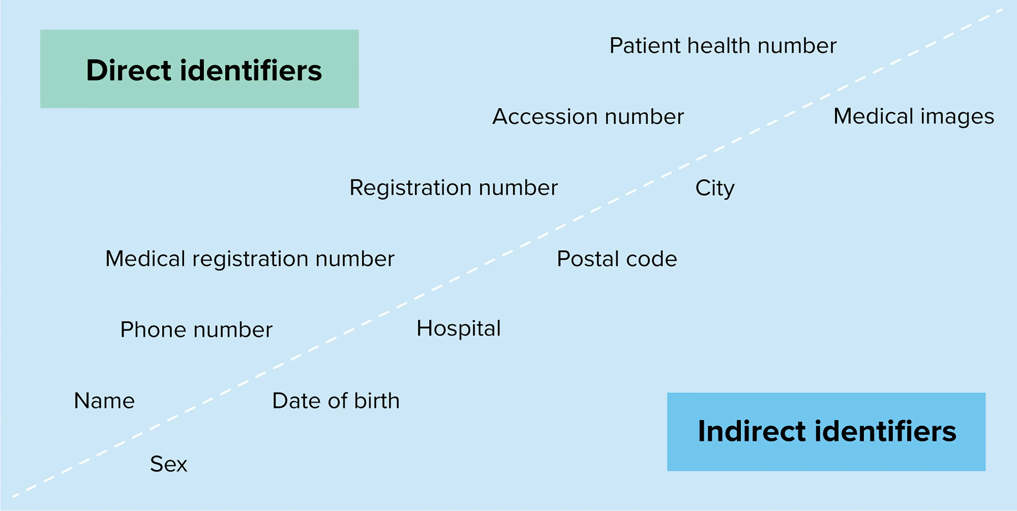
4) Data Security and Privacy: Medical images and protected health information (PHI) must be safeguarded against unauthorized access, breaches, and cyber threats to maintain patient confidentiality.
Key measures include:
- Robust Security Protocols: Implementing industry-standard encryption, secure authentication mechanisms, and secure network protocols to protect data during storage and transmission.
- Regulatory Compliance: Adhering to regulations such as the Health Insurance Portability and Accountability Act (HIPAA) in the United States and the General Data Protection Regulation (GDPR) in the European Union is essential. Compliance ensures that patient data are managed according to strict confidentiality and privacy standards.

By understanding these aspects, we aim to emphasize the core principle that, when leveraged thoughtfully, data can lead to powerful, efficient, and robust AI advancements in the field of radiology.
What are the next steps? As we stand on the brink of this revolutionary shift, healthcare providers, legislators, and innovators need to come together more frequently and in greater numbers. Understanding the legal landscapes and preparing for the changes they bring will ensure that we harness AI's full potential responsibly and effectively.
Do you want to delve deeper into how these technologies are shaping the future of radiology and healthcare? Click here to learn more about current AI vendors in Radiology.


Discover how Rayscape works
integrate Rayscape AI into
your hospital's infrastructure
and improve both your
daily workflow and patients' experience.
