

How AI Platforms Improve Radiology Workflow Optimization in 2025
Artificial intelligence (AI) is increasingly revolutionizing the field of radiology by enhancing workflow efficiency and productivity beyond mere image interpretation. AI-powered tools are automating routine administrative tasks such as data entry and image sorting, which allows radiologists to devote more time to patient care and the complex nuances of image analysis.
In today's medical environment, characterized by escalating workloads, more intricate scans, and the necessity to reduce costs and minimize errors, these AI applications have become indispensable. This article provides a comprehensive overview of the current advancements in AI applications that significantly impact radiology workflows.
Enhancements in Image Acquisition Through AI
The foundation of accurate medical image interpretation lies in the quality of image acquisition. Various factors—including radiation dose, image resolution, patient movement and positioning, implanted medical devices, and sensor inconsistencies—can influence the quality of the images obtained. Research indicates that machine learning (ML) techniques in this area can reduce radiation exposure, shorten scanning times, decrease false-positive rates, and minimize unnecessary repeat imaging, all while preserving image integrity.
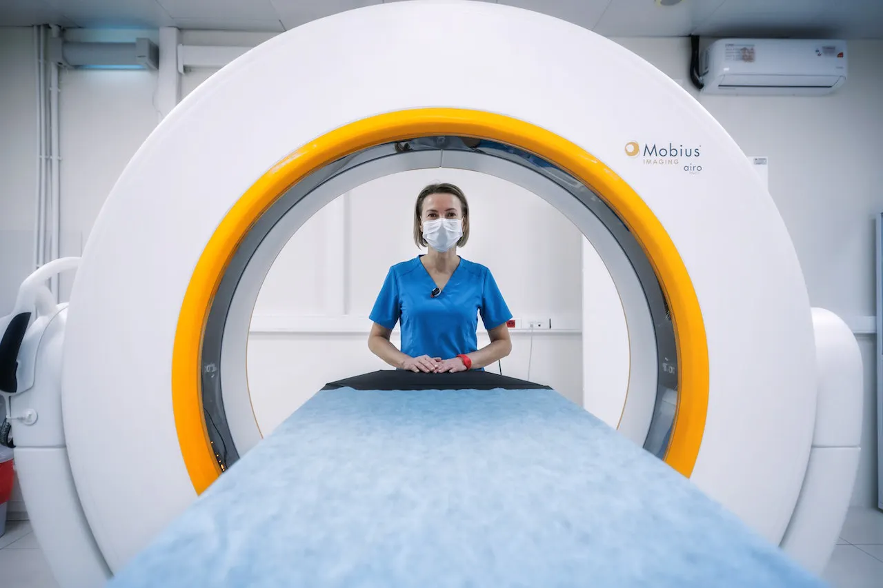
Reducing Radiation Dose
With the global rise in the utilization of computed tomography (CT) and positron emission tomography (PET) scans, concerns about patient radiation exposure have intensified, particularly for those undergoing frequent imaging. Radiology departments often grapple with balancing image quality maintenance against the imperative to minimize radiation doses, adhering to the principle of keeping exposure "as low as reasonably achievable." Traditionally, reducing radiation dose in CT scans involves lowering the tube current, a method that, while effective, can introduce noise and reduce diagnostic confidence.
Recent advancements in AI-driven image reconstruction offer promising alternatives. These techniques enhance the quality of low-dose CT images, making them comparable in clarity to images acquired at standard doses. Deep learning models are trained on datasets comprising both low-quality, noisy images and high-quality counterparts, enabling them to recognize pathologies and normal structures even at reduced doses. As a result, low-dose images can be reconstructed to appear as if they were obtained with regular radiation levels.
Advancements in Image Reconstruction
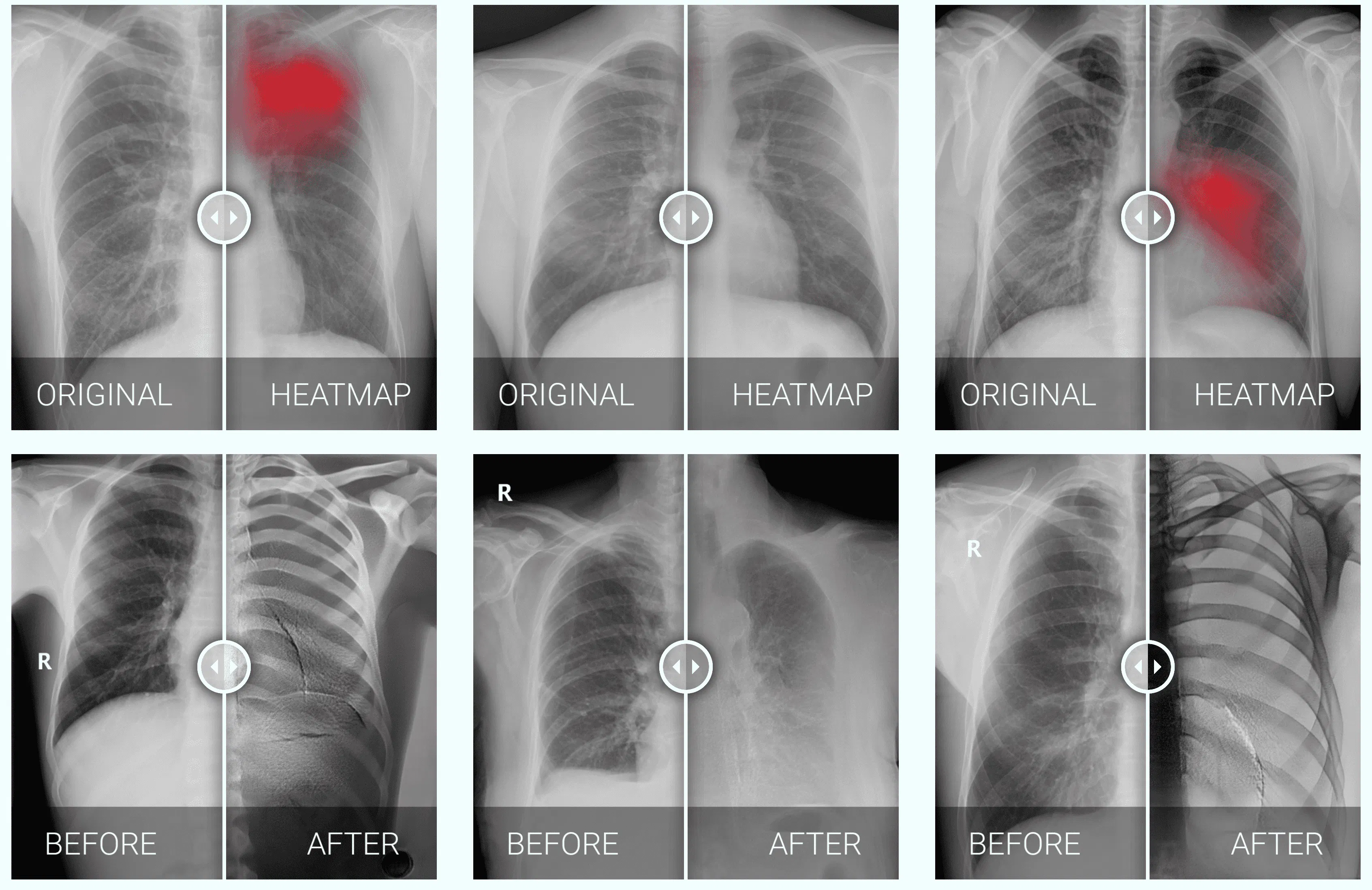
Image reconstruction is a critical component in medical imaging, aiming to produce high-quality diagnostic images while managing costs, reconstruction time, and patient safety. Extensive research has focused on leveraging ML techniques to improve image reconstruction across CT, magnetic resonance imaging (MRI), and PET scans. Key targets for enhancement include noise reduction, artifact suppression, motion compensation, accelerated image acquisition, and multimodal image registration. These objectives are interconnected, often enabling the reduction of both radiation and contrast agent doses through the successful implementation of advanced reconstruction methods.
Deep learning solutions have been developed to:
- Denoise CT and MRI images without sacrificing technical details.
- Reduce noise and suppress artifacts in MRI, thereby shortening acquisition times.
- Reconstruct PET images obtained with low-dose radiotracers, making them resemble images produced with full-dose radiotracers.
Improving Patient Positioning
Patient positioning presents several challenges in radiology. Accurate positioning is vital for precise imaging and diagnosis but can be hindered by factors such as patient discomfort, mobility limitations, or lack of cooperation. Incorrect or inconsistent positioning may lead to suboptimal image quality, necessitate repeat scans, increase radiation exposure, and delay diagnoses. Additionally, certain required positions, such as those in mammography, can be physically uncomfortable or anxiety-inducing for patients, complicating the acquisition of high-quality images.
Recent AI advancements in this area include:
- In CT imaging: Solutions utilizing 3D depth-sensing cameras that recognize anatomical landmarks and automatically calculate the patient's center, optimizing bed positioning for dose efficiency and image quality.
- In mammography: Systems that automatically assess image quality at the time of acquisition to ensure compliance with relevant quality standards and regulations, such as the Mammography Quality Standards Act (MQSA) and international guidelines.
Optimizing Worklist Prioritization
Radiologists' worklists are traditionally populated based on predefined criteria like the body part to be imaged, imaging modality, patient location, and examination urgency. Issues arise when non-emergency exams are incorrectly flagged as urgent to expedite the imaging process. This practice can blur the lines between routine and emergency cases, potentially delaying the interpretation of truly critical examinations.

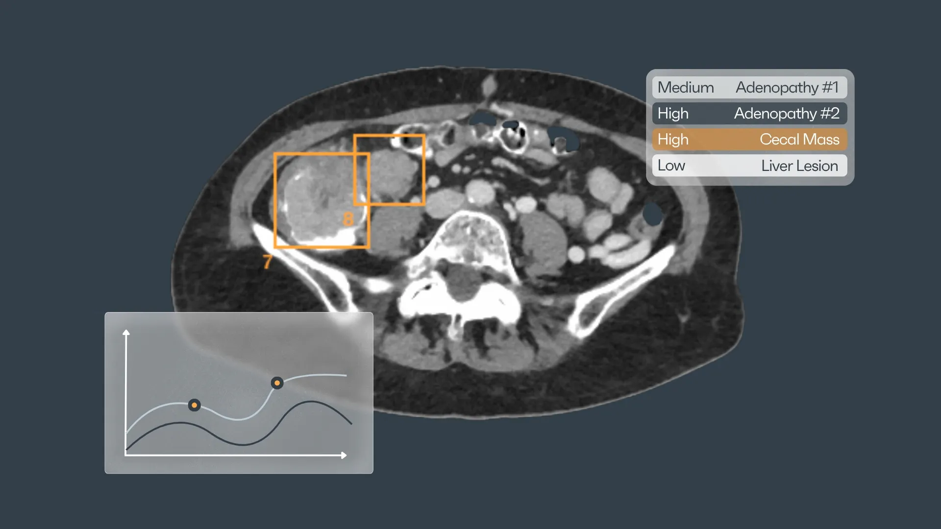
To address this problem, AI algorithms have been developed to prioritize examinations with emergent findings across multiple body regions. These models must maintain high sensitivity and specificity to detect urgent cases while minimizing the occurrence of false positives, ensuring that genuinely urgent cases receive prompt attention.
Streamlining Study Protocoling with AI
Protocoling involves selecting appropriate sequences for MRI or CT scans to accurately capture the desired anatomical structures and abnormalities. Typically performed by radiologists due to their specialized expertise, this process requires reviewing clinical data and ordering information from electronic health records, relevant lab values, and prior images and reports. This task is time-intensive, often consuming one to two hours daily in each radiology department sector.
AI offers significant benefits in developing study protocols and customizing hanging protocols for individual radiologists. While most Picture Archiving and Communication Systems (PACS) provide automatic hanging protocols, they are generally standardized, displaying images similarly for all users. Radiologists often have unique display preferences. AI-driven software solutions are evolving to adapt to these preferences, arranging images accordingly. This area is particularly suited for AI applications because inconsistencies in DICOM metadata and varying sequence names used by different vendors make traditional rule-based algorithms less effective for optimizing hanging protocols.
Enhancing Study Reporting
Integrating AI into radiology reporting can improve the clarity, accuracy, and quality of reports while reducing variability. There is growing interest in deploying deep learning natural language processing models that act as smart assistants or automate tasks such as populating recommendations for follow-up on incidental findings. These applications can streamline the reporting process, ensuring consistency and aiding radiologists in generating comprehensive reports.

Integrating AI into Medical Education
With artificial intelligence increasingly infiltrating the medical profession, it is imperative that trainees acquire foundational knowledge in AI—a need highlighted by studies showing their enthusiasm for such learning. Regardless of whether they specialize in radiology, AI will play a significant role in their future practices. AI-powered tools are now assisting not only in image interpretation but also in ordering appropriate studies and managing patient follow-up. As educators, it is our responsibility to equip these future professionals with a solid understanding of the tools they will utilize, building a foundation upon which they can expand.
Despite its vast potential, AI is not without flaws, making it essential to critically assess the outputs generated by these systems. Trainees must master the basics to effectively interpret the information provided by AI tools and to make informed decisions about their trustworthiness. Physicians, with their extensive training, remain the domain experts, and AI systems will never fully replicate the intangible qualities—the ability to think beyond conventional boundaries—that doctors possess.
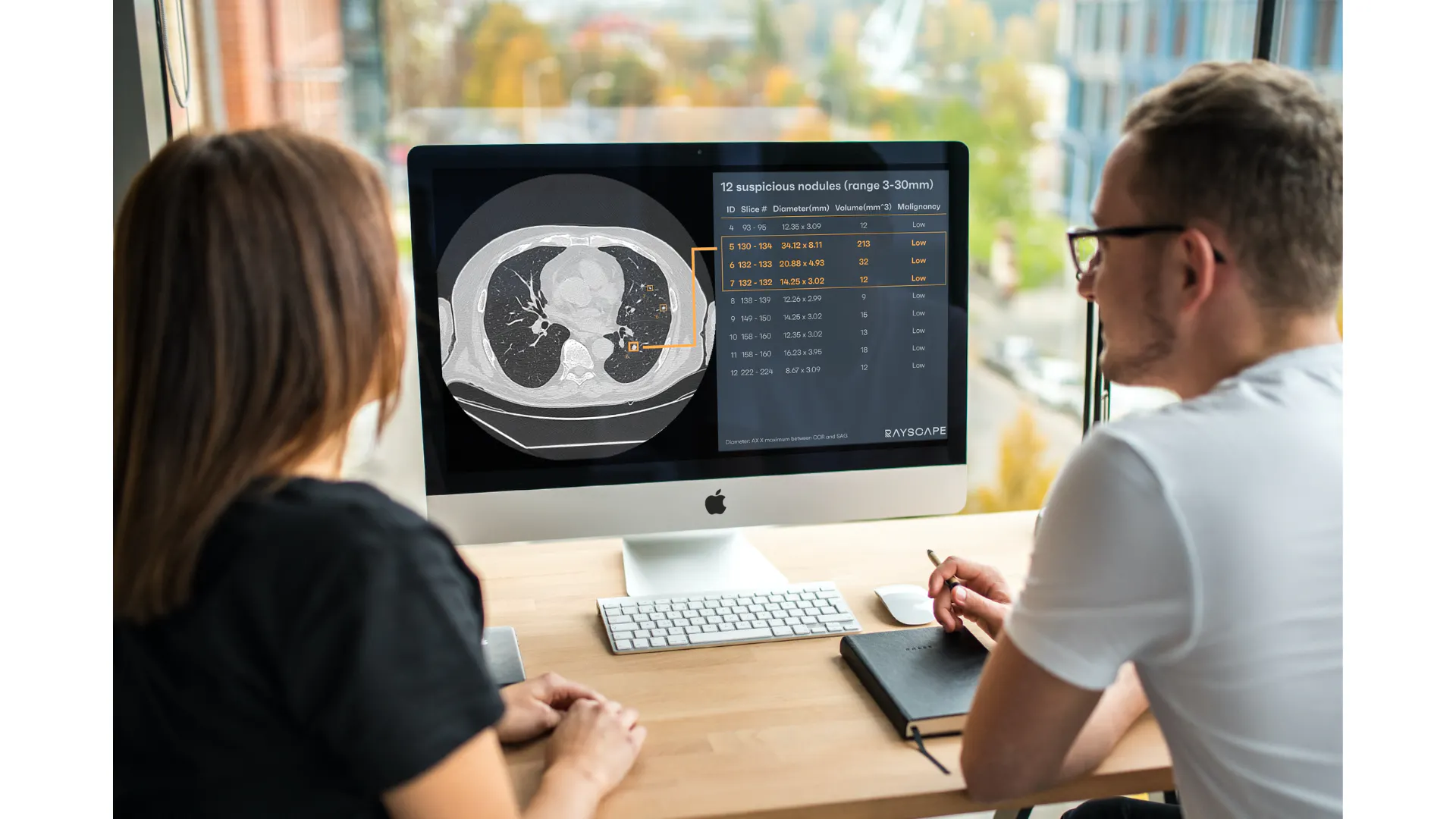
Incorporating AI into medical education prepares upcoming healthcare professionals for the shifting terrain of medicine. AI can grant students access to extensive datasets and a multitude of case examples, thereby enhancing their exposure to a wide range of pathologies and enriching their learning experiences. Through AI-driven platforms that simulate real-life scenarios, students can practice interpreting and diagnosing medical images, accelerating their skill development. AI algorithms serve as valuable decision-support tools, aiding radiology students in analyzing complex images and detecting subtle anomalies. By weaving AI into radiology training, students can bolster their diagnostic precision, build confidence, and become more adept at identifying and assessing various conditions.
Just think about this: AI can automate labor-intensive tasks like image segmentation and measurement, allowing radiology trainees to focus on more critical aspects of their education. This boost in efficiency can streamline workflows and increase productivity, affording residents additional time to learn and hone their diagnostic skills. AI platforms are also capable of adapting to individual learning needs, offering customized educational content and feedback. By evaluating students' performance and pinpointing areas needing improvement, AI can tailor educational modules to address specific learning gaps, ensuring personalized learning journeys.
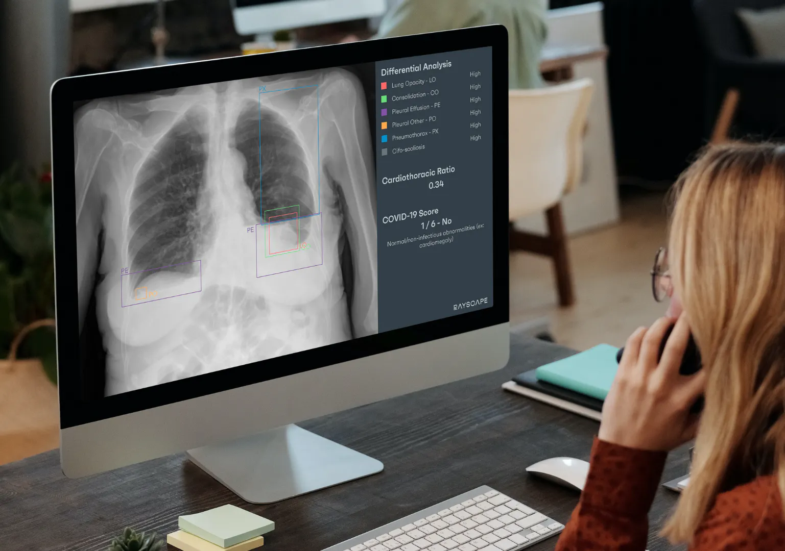
Trainees versed in AI could contribute to the development of AI-driven clinical decision-support systems, aiding in the formulation of personalized treatment plans based on patient-specific data. A deep understanding of AI's technical aspects empowers trainees to critically appraise and validate AI algorithms, ensuring their proper application in patient care. This proficiency not only enhances their clinical capabilities but also opens doors to new opportunities within the industry, as these skills are highly sought after. Ultimately, a thorough grasp of AI enables medical trainees to embrace the future of medicine, contributing to progress in diagnostics, treatments, and patient outcomes.

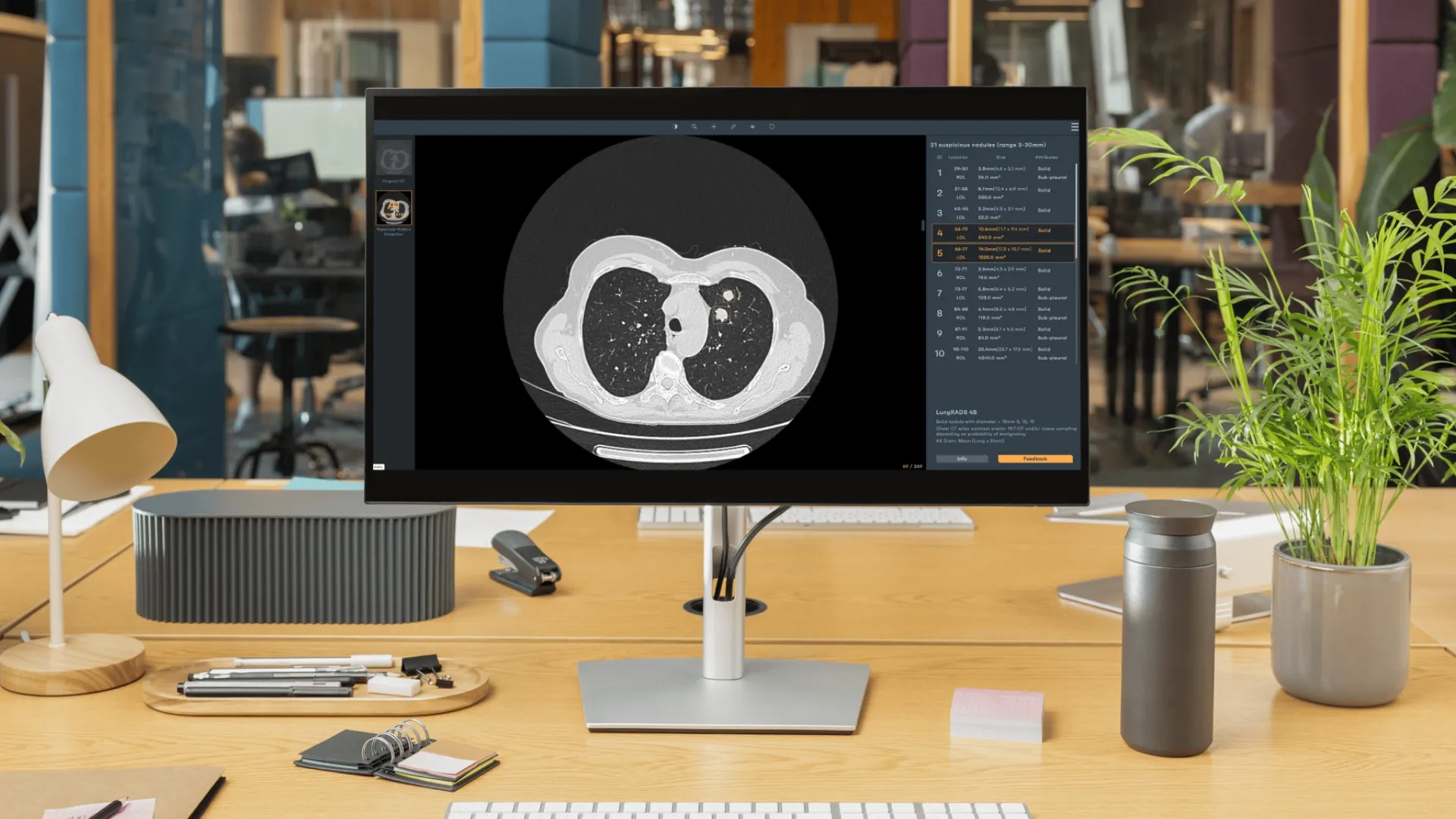
🩻In recent years, we've witnessed incredible advancements in technology, but we know that the real challenge is bringing these innovations directly to you -𝐭𝐡𝐞 𝐫𝐞𝐚𝐥 𝐮𝐬𝐞𝐫𝐬- in a way that feels seamless and truly impactful. Our new web demo app is designed with this goal in mind—𝐛𝐫𝐢𝐝𝐠𝐢𝐧𝐠 𝐭𝐡𝐞 𝐠𝐚𝐩 𝐛𝐞𝐭𝐰𝐞𝐞𝐧 𝐩𝐨𝐭𝐞𝐧𝐭𝐢𝐚𝐥 𝐚𝐧𝐝 𝐩𝐫𝐚𝐜𝐭𝐢𝐜𝐚𝐥 𝐚𝐩𝐩𝐥𝐢𝐜𝐚𝐭𝐢𝐨𝐧, and putting powerful features directly in your hands for testing.
🖥️ 𝐍𝐨 𝐜𝐨𝐧𝐭𝐫𝐚𝐜𝐭𝐬 𝐧𝐞𝐞𝐝𝐞𝐝, 𝐭𝐨 𝐭𝐞𝐜𝐡𝐧𝐢𝐜𝐚𝐥 𝐢𝐧𝐭𝐞𝐠𝐫𝐚𝐭𝐢𝐨𝐧, 𝐧𝐨 𝐩𝐚𝐲𝐦𝐞𝐧𝐭 𝐜𝐨𝐦𝐦𝐢𝐭𝐦𝐞𝐧𝐭
Try Rayscape Webdemo Today at: https://demo.rayscape.ai/home
At Rayscape, we stand committed to pushing the boundaries of radiology management software, integrating the power of artificial intelligence to revolutionize patient care, enhance efficiency, and drive unprecedented advancements in the field of radiology. Join us on this transformative journey, where innovation meets excellence to shape the future of radiology.


Discover how Rayscape works
integrate Rayscape AI into
your hospital's infrastructure
and improve both your
daily workflow and patients' experience.
