

Artificial Intelligence's Impact on Pulmonology in 2025
In the realm of pulmonary medicine, the early detection and characterization of Incidental Pulmonary Nodules (IPNs) are paramount for enhancing patient outcomes. IPNs, often the initial indicators of potential lung cancer, have traditionally been identified through manual reviews or keyword searches of chest X-ray (CXR) and CT scan reports by radiologists or pulmonologists.
Human oversight is inevitable, and subtle nodules can be missed, leading to delayed diagnoses and treatments. This is where Artificial Intelligence (AI) emerges as a revolutionary force, offering powerful tools for early IPN detection and reshaping the landscape of thoracic medicine.
By harnessing extensive datasets of chest X-rays, AI algorithms have been trained to detect clinically significant IPNs with exceptional accuracy, consistency, and high sensitivity and specificity. These advanced systems can identify suspicious nodules that human observers might overlook—in some instances, up to 70% of radiographs—substantially increasing the overall detection rate and broadening the scope for early lung cancer identification. Early detection through AI enhances the pulmonology diagnostic funnel, enabling interventional pulmonologists to initiate diagnostic bronchoscopies sooner, thereby accelerating the path to definitive diagnosis and treatment.

The implications of AI-assisted early IPN detection are profound. By swiftly integrating patients into investigative pathways, surgeons, radiologists, and pulmonologists can collect tissue samples for cancer cell analysis at earlier stages. This expedited process facilitates quicker conclusive diagnoses, allowing patients to enter appropriate treatment pathways more promptly, potentially even leading to curative outcomes. Undoubtedly, AI is a game-changer in the quest for early intervention and improved survival rates from the world's deadliest cancer.
Optimizing Medical Device Innovations Aligned with AI
Pulmonology has witnessed a remarkable transformation with the advent of robotic bronchoscopy. This innovative technology combines the precision of robotics with the flexibility of traditional bronchoscopy, enabling pulmonologists to conclusively diagnose lung cancer at earlier stages. Utilizing thin, maneuverable catheters guided by advanced robotics, physicians can now access peripheral lung nodules that are unreachable by CT-guided biopsy, achieving unparalleled accuracy and stability. This minimally invasive approach not only enhances diagnostic yields but also significantly reduces the time from initial nodule detection via X-ray or CT to biopsy, allowing patients to receive timely treatment. The technical advancements and clinical implications of this groundbreaking technology are poised to revolutionize the early detection and management of lung cancer.
Leveraging AI to Boost Clinical Confidence and Efficiency
Beyond its role in early detection, AI offers numerous benefits that bolster clinical confidence and streamline workflows in pulmonology. One key advantage is the standardization of nodule assessment. AI ensures consistent evaluation of chest X-rays, mitigating inter-observer variability among radiologists and pulmonologists while upholding high diagnostic accuracy. This standardization enhances the reliability and reproducibility of IPN detection across different healthcare settings.
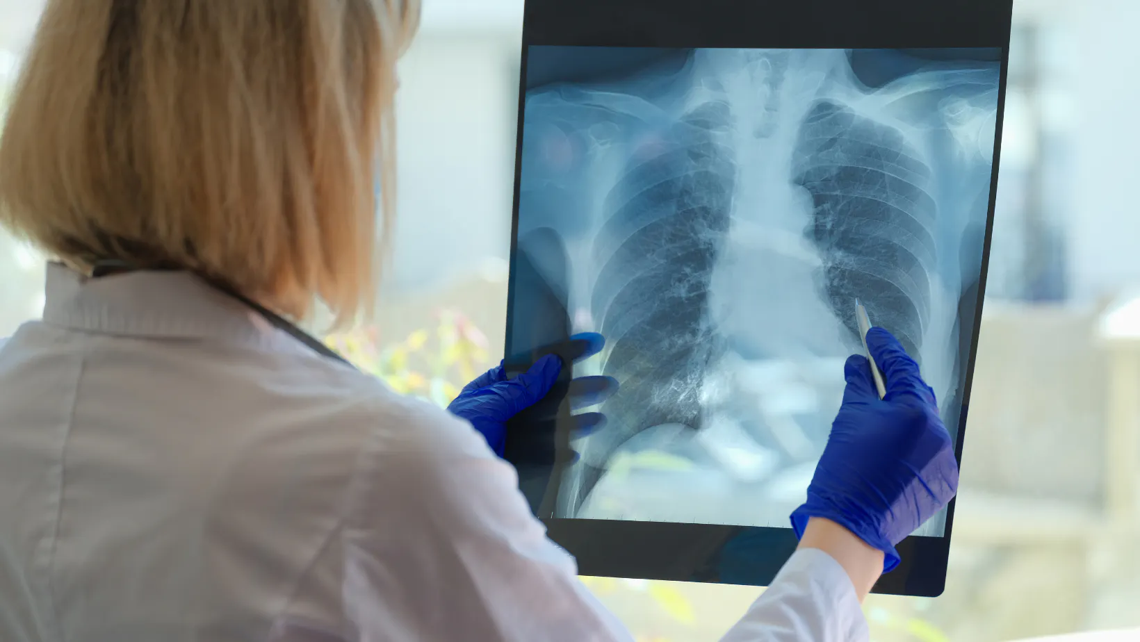
Furthermore, AI's rapid ability to identify IPNs accelerates subsequent diagnostic tests and interventions, hastening patient access to care. This efficiency optimizes clinical workflows and enables pulmonologists to make informed treatment decisions more rapidly. The seamless integration of AI into the diagnostic process saves time and allows for a more streamlined approach to patient management.
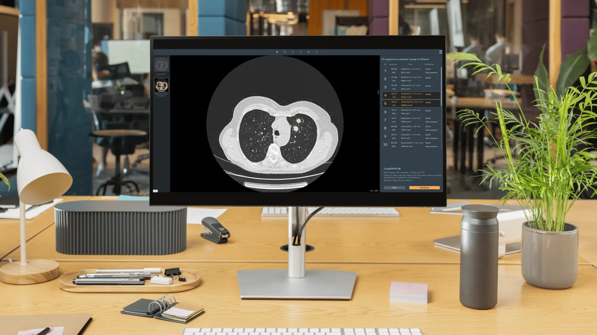
An additional significant aspect of AI's impact on clinical confidence lies in its advanced nodule analysis capabilities. AI can generate Lung Nodule Malignancy Risk Scores, aiding in differentiating between benign and malignant nodules. This sophisticated analysis reduces false positives and unnecessary procedures while providing invaluable clinical decision support. The risk scores incorporate nodule characteristics such as texture, composition, and calculations of volume doubling time.
AI also helps alleviate patient anxiety by accurately and promptly identifying nodules that require further intervention—and importantly, those that do not warrant biopsy. Integrating AI-powered nodule analysis empowers pulmonologists to make more precise and informed decisions, ensuring patients receive the most appropriate care based on their individual needs.
AI-generated reports offer detailed insights and visual aids that facilitate effective communication between pulmonologists and patients. These comprehensive and organized reports improve patient understanding and engagement in their care plans, fostering a patient-centric approach. The synergy of technological innovation and human expertise creates a collaborative environment where optimal outcomes can be achieved.
Envisioning the Future of Pulmonology Through AI
The integration of AI into pulmonology pathways holds immense promise for optimizing patient care. Early lung cancer detection through AI-assisted IPN identification can significantly improve survival rates. Moreover, AI's ability to enhance clinical decision-making, guide interventions, and optimize workflows has the power to fundamentally transform the practice of pulmonology.
As the field of AI continues to evolve, innovative solutions like Rayscape CXR are at the forefront of harnessing artificial intelligence to revolutionize pulmonary medicine. By embracing these cutting-edge technologies, pulmonologists can usher in a new era of precision, efficiency, and patient-centered care.
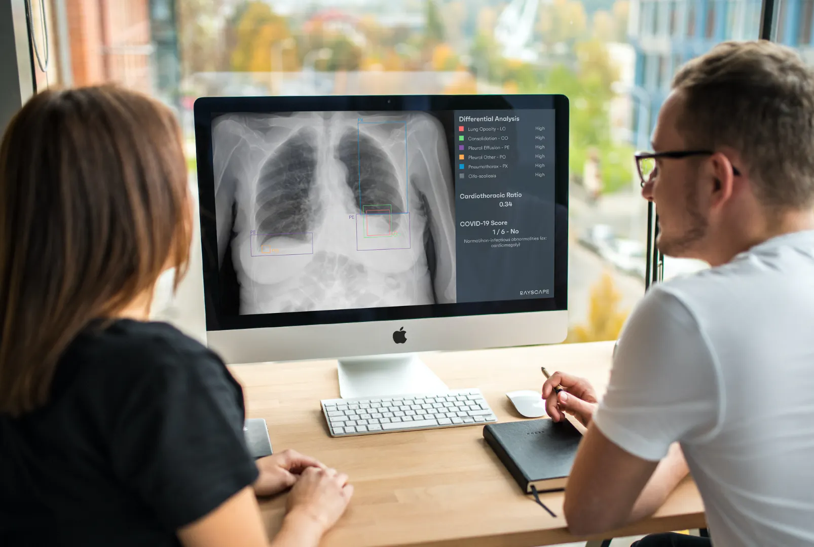
The digital future of pulmonology has arrived, and AI is poised to make a significant impact in the fight against lung cancer. With its capabilities in early IPN detection, enhancing clinical confidence, guiding bronchoscopy interventions, and improving patient outcomes, AI stands as a pivotal tool. As we stand on the cusp of this transformative era, it is an exciting time where innovation and dedication converge to shape a brighter, healthier tomorrow.
Resources:
1. World Health Organization (WHO)
Cancer Fact Sheet.
Link: https://www.who.int/news-room/fact-sheets/detail/cancer
2. Ardila, D., Kiraly, A.P., Bharadwaj, S., et al. (2019)
End-to-end lung cancer screening with three-dimensional deep learning on low-dose chest computed tomography.
Nature Medicine, 25(6), 950–958.
Link: https://www.nature.com/articles/s41591-019-0447-x
3. European Society of Radiology (ESR) and European Respiratory Society (ERS) (2015)
Lung cancer screening: recommendations.
European Respiratory Journal, 46(1), 28-39.

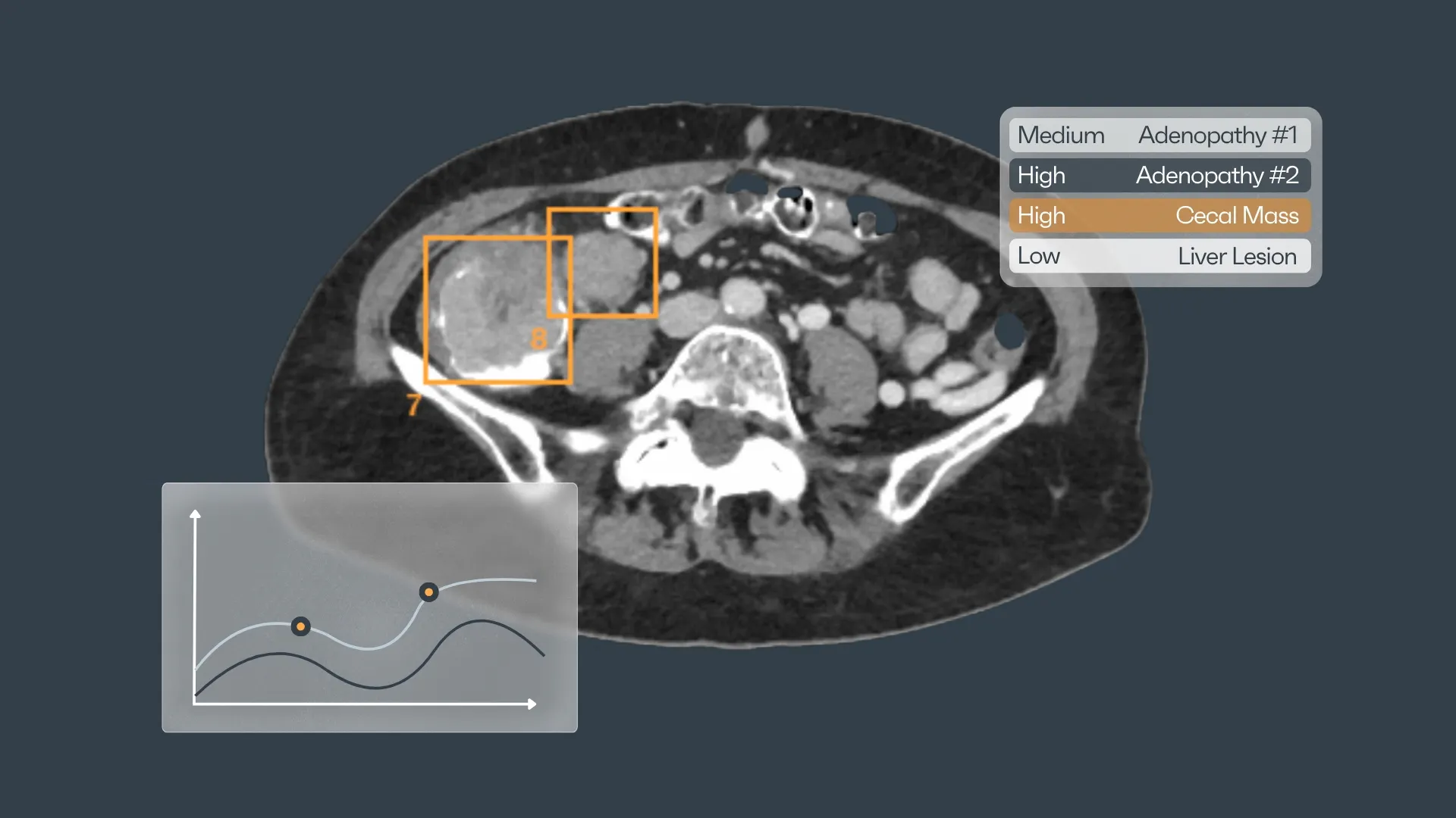

Discover how Rayscape works
integrate Rayscape AI into
your hospital's infrastructure
and improve both your
daily workflow and patients' experience.
