

How to Implement AI in Radiology Workflows: A Practical Guide
Before we begin, let us clarify: this is not a story about a machine that changed everything overnight. There is no dramatic breakthrough, no singularity concealed behind a press release. Instead, this is the story of a hospital—an academic medical center in the Netherlands—methodically teaching itself how to live with artificial intelligence.
Let’s call it “Southern” as the researchers did. A pseudonym, yes, but one that now carries the weight of real-world progress. Southern is not just a site on the map but a symbol of what healthcare systems across Europe are quietly grappling with: how to implement AI meaningfully, sustainably, and at scale.
Radiology, by nature, has always had an affinity for abstraction. It interprets shadows, reads meaning from patterns, decodes the invisible. In many ways, it was destined to be the first field in medicine to invite AI into the room. And so, between 2020 and 2025, more than half of all CE-marked medical AI tools were designed for radiology. Interest surged, as did investment. At RSNA2024 and ECR2025, the number of AI vendors nearly doubled from the year before, a number that was also double from the previous year. Yet, clinical adoption still remains slow.
Despite a wave of innovation, studies repeatedly cite similar obstacles: fragmented IT infrastructure, lack of automated data routing, limited scalability of narrow AI tools, poorly defined clinical use cases, and a pervasive uncertainty among both clinicians and managers on where—and how—AI should be deployed. It is against this backdrop that “Southern” offers a rare and instructive glimpse into what real-world implementation can look like: complex, iterative, and above all, human.
For a period of 4 years, a team of researchers conducted a longitudinal qualitative case study at Southern. Over the course of 43 non-consecutive observation days, they attended 30 internal meetings, conducted 18 in-depth interviews, and collected 41 documents related to AI projects underway within the radiology department. Southern was selected precisely because of its position as an early mover in the field. At the time of the study, the medical center was running more than 15 AI implementation projects across radiology, making it one of the most active institutions of its kind in Europe.
This wasn’t a theoretical inquiry. The research aimed to extract actionable, transferable insights that could support other healthcare organizations facing similar challenges. Using abductive reasoning—a flexible analytical approach that moves between data and theory—the team coded the AI-related change initiatives at Southern and identified how they addressed (or failed to address) known barriers to implementation.
What emerged were three core thematic domains that structure the AI transformation at Southern: technological infrastructure, clinical workflow integration, and organizational change.
1. Building the Foundation: Technological Infrastructure
One of the most significant barriers to AI deployment in clinical settings is the complexity of hospital IT ecosystems. AI models may function impressively in isolated settings, but in the wilds of real clinical environments, they often stumble. Southern tackled this head-on. Their approach wasn’t simply to “install” AI but to actively prepare the environment around it. This included upgrading data routing systems, ensuring proper DICOM formatting, enabling seamless integration with PACS, and aligning local compute resources with the demands of inference-time processing.
Rather than forcing AI tools into existing frameworks, Southern adapted its infrastructure to accommodate them—a strategy akin to preparing the soil before planting. These were not superficial fixes but deep structural changes that reflected a long-term commitment to technological readiness. And, crucially, the hospital’s IT and clinical teams collaborated from the outset. AI wasn’t treated as a siloed research project but as an operational priority that would only succeed through cross-disciplinary alignment.
2. Designing for Use: Workflow Integration
The second challenge Southern confronted was not technological, but behavioral. Radiology is not just a series of clicks—it’s a ritual. A cadence. AI, to be accepted, has to become invisible. Seamless. It could not be another window to monitor, another platform to switch into. So Southern embedded AI into the flow of work the same way water finds cracks in stone: slowly, persistently, without seeking permission. Integration became an art. Interfaces were tailored. Outputs were streamlined. The goal was not just accuracy—it was familiarity. One observed project used AI to flag potential anomalies in imaging studies. But the success wasn’t just in detection rates—it was in how and when those flags appeared. Were they disruptive? Were they ignored? Over time, AI became less of a novelty and more of a quiet assistant: present, reliable, and unobtrusive.
3. Culture Eats Code: Organizational Transformation
The most complex challenges were not technical but cultural. Implementing AI in radiology required more than software licenses and integration protocols—it demanded a new mindset. The team recognized early that adoption would depend on buy-in from clinicians, managers, and support staff alike. To that end, they created formal governance structures, including steering committees and designated AI leads within departments. They developed educational materials tailored to different roles, helping non-technical stakeholders understand not only how the algorithms worked but why they were being introduced. Perhaps most importantly, they framed AI as a tool for augmentation, not automation. Radiologists were still the final interpreters, still the authors of reports. But now, they had a new colleague in the background: one trained not in years, but in datasets. This emphasis on trust and transparency made all the difference.
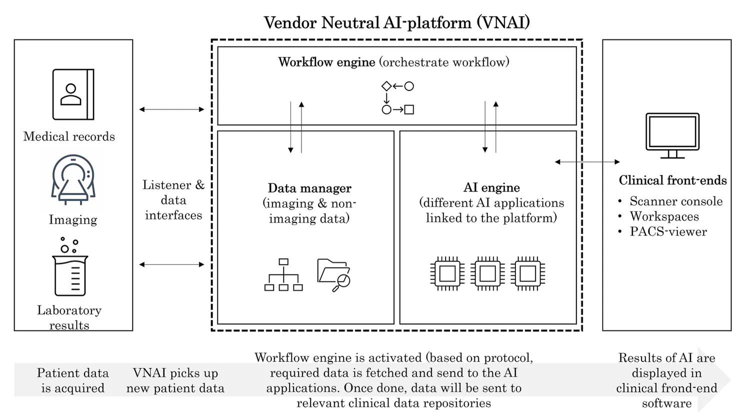
Fig. 1 Hospital-level vendor-neutral AI (VNAI) platform towards a scalable and seamless AI implementation
Where the Machine Begins: Southern’s Response to the Real-World Limits of AI
The first thing we need to understand is this: Artificial intelligence, in its current clinical form, is a specialist. A narrow-functioned, single-task expert—fast, focused, and frustratingly limited. It does one thing well. Maybe it segments lungs. Maybe it flags nodules. Maybe it whispers that something doesn’t look quite right. But it doesn’t do everything. Therein lies the problem. Radiologists don’t deal in single-task days. Their work is layered—an orchestration of impressions, modalities, histories, anomalies, and timing. They don’t want a chorus of tools interrupting them mid-thought.
They want harmony. Flow. Trust.
Long before many others did, doctors from Southern recognized that successful AI deployment would require not just the right tools, but the right infrastructure to host them. To let them speak to each other. To let them disappear into the background of daily use.
The Invisible Conductor: Southern’s Vendor-Neutral AI Platform
Imagine, if you will, a kind of quiet conductor—an unseen intelligence behind the scenes, routing images, sorting protocols, guiding each scan toward the algorithm best suited for its analysis. That’s Southern’s vendor-neutral AI platform, or VNAI. And while its name may sound like bureaucracy in disguise, its function is anything but.
The VNAI is, in many ways, a nervous system for the hospital’s AI ecosystem. It doesn’t do the diagnosis—but it makes sure the right tool is called into play at the right time, for the right scan, in the right sequence. No user prompts. No manual data pushes. Just metadata—modality, protocol, patient age—and silent orchestration. This was Southern’s answer. Not a flash of intelligence, but a slow, methodical calibration of timing. A rethinking of when and how AI appears inside clinical practice. A commitment to invisibility, where the system’s presence is measured not in novelty, but in the absence of friction.
A Chorus of Vendors, A Single Channel
Here’s the reality most hospitals face: every AI vendor arrives with their own terms, their own formats, their own paperwork. Each algorithm is a guest with its own dietary needs, legal quirks, and incompatible plugs. The VNAI offered a solution not just to workflow, but to regulation, security, and cost. By centralizing the legal and technical framework, they reduced the redundancy of reviewing every application in isolation. One security review. One data flow. One master agreement.
From a logistical perspective, the VNAI didn’t just simplify deployment—it accelerated it. Before the platform, implementing a single AI solution often took over a year. Not because the software was complicated, but because the institutional scaffolding around it—the legal documents, the integration plans, the ethical reviews—moved at a glacial pace. With the VNAI in place, new applications could go live in a matter of weeks.
This isn’t to say the technology alone solved everything. Multiple other forces—vendor maturity, staff availability, internal expertise—contributed to acceleration. But the VNAI became a kind of multiplier. A scaffold upon which multiple applications could coexist. Importantly, only certified AI applications were hosted on the platform. This allowed for rapid testing of solutions that were already cleared for clinical use—transforming Southern from a passive adopter into an agile evaluator. Algorithms could be deployed, observed, iterated upon—and if needed, removed—with minimal disruption. And then, in another quiet but crucial move, the teams shifted the VNAI from an on-premise setup to a cloud-based infrastructure. The implications were enormous: easier updates, fewer maintenance demands, and a scalable architecture that could grow with the field.
One Click Too Many: The Interface as Gatekeeper
There is a kind of violence in too many clicks. Not literal, of course, but cognitive. When a clinician has to break focus to open a separate tool, launch a browser, wait for results, copy-paste findings—that’s not augmentation. That’s interruption. Rather than settle for superficial integration—say, a hyperlink that opens an external dashboard—they pushed for seamless UI design. AI outputs had to appear in the same viewer radiologists already used: PACS. Advanced image viewers. Whatever the clinician was already familiar with—that’s where the AI would live.
This meant co-creating with vendors. Customizing APIs. Requesting outputs that didn’t just make sense to machines, but to human users in a hurry. And in a rare moment of consensus across clinical and managerial lines, everyone agreed: seamless integration was non-negotiable. Not because it was easier to sell, but because it was essential to use. A frictionless interface was not a luxury—it was a survival mechanism for adoption.
If the VNAI was the invisible spine of Southern’s AI strategy, then its nervous system—the thing that moved, adapted, and responded—was the workflow. Not code. Not compute. But practice. You might imagine AI slides neatly into clinical routines, like a new app on your phone or a faster processor in your laptop. But hospitals don’t work like that. Medicine isn’t software. It’s lived. It's layered. It's institutional memory and handoffs and deeply ingrained ritual. You can't just drop an algorithm in and expect the machine to hum.
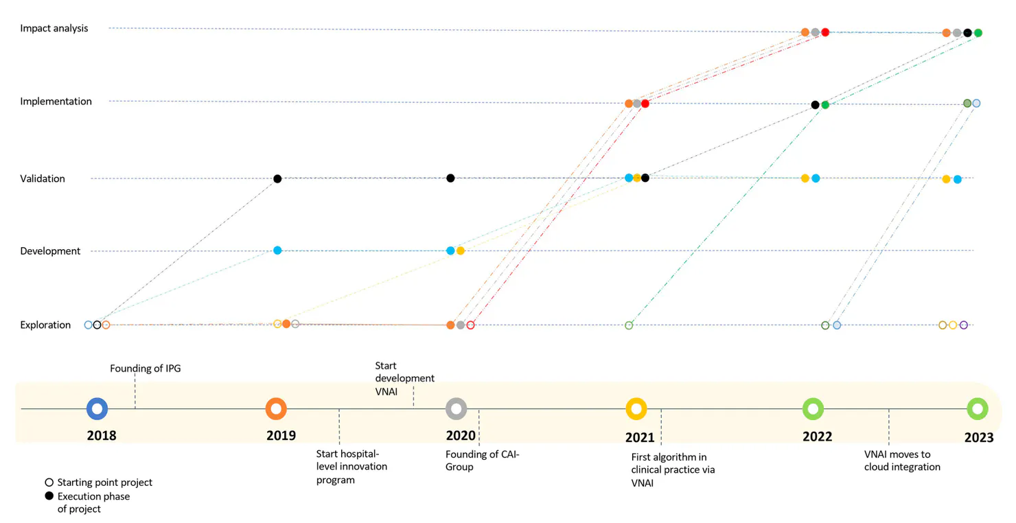
Fig.2: Overview of progress of AI projects over time. Open dots represent the starting point of the project, whereas solid dots represent the progress of the projects over time. Important events are presented on the timeline at the bottom of the figure.
The Image Processing Group: A New Kind of Center
Somewhere inside the radiology department, behind doors that don't bear the word “AI” but probably should, sits the Image Processing Group (IPG). It was formed not as a gesture to innovation, but as a response to a mounting tension: AI tools were being bought, built, installed… and then left unused. Not out of resistance, but out of rhythm. “Although costing a fortune,” said the department chair, “image analysis tools were not being used in a consistent manner.”
So they created the IPG—a living, breathing hub staffed not by futurists or engineers, but by radiographers and technical physicians. Nine of the former, two of the latter. Their role? To ensure that technology doesn’t just sit in the system but moves through it. That it becomes habit. That it works. They pre-populate reports, standardize protocols, and make sure the AI’s contribution arrives not as noise, but as signal. In doing so, they redefine what it means to “read” an image. The AI does its part. The radiographer does theirs. And the radiologist—still the final voice—receives something closer to clarity.
We must understand that AI doesn’t always travel well. A model trained on one dataset might falter in a new environment—a different patient population, different scanner, different disease prevalence. What worked in India or the United States may not work in Poland. So the IPG’s technical physicians became translators—fluent in both engineering and medicine. They didn’t just validate AI models once. They continuously tested them, monitored them, asked questions the algorithms couldn’t answer on their own. They ran a retrospective study on over 15,000 chest X-rays to see if an AI application built elsewhere could spot the abnormal from the normal, accurately and reliably, here. Then they built a dashboard—not to celebrate success, but to track alignment between AI results and actual clinical reports. It became a mirror. A measure of consistency. A safeguard.
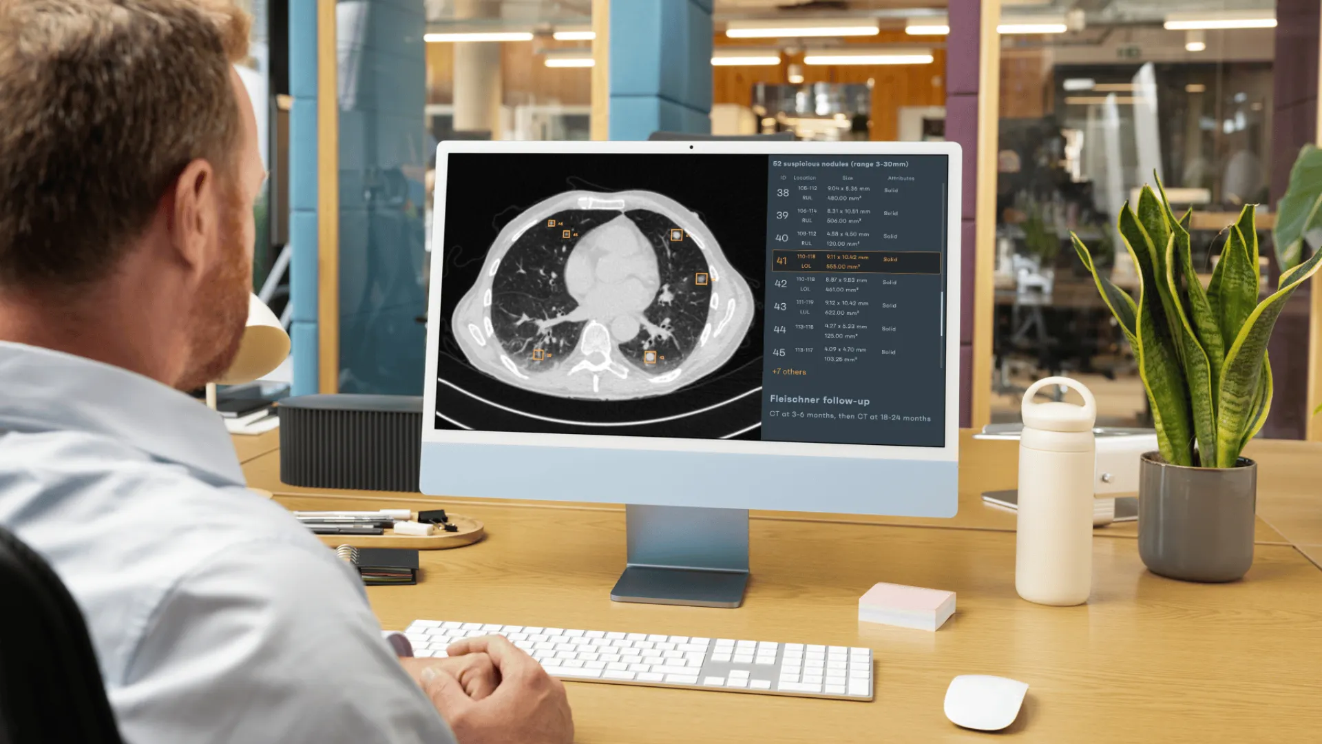
One of the most telling insights from Southern is this: clinicians will not trust what they cannot change. Many AI outputs come fixed, rendered as PDFs or still images, frozen in form. But not for diagnosis. Not for nuance. Radiologists want to engage with results, not just observe them. So when the team implemented an AI solution for lung nodule detection on CT, they refused to accept locked outputs. Instead, they negotiated—with both the AI and PACS vendors—to make the results modifiable inside the PACS viewer itself.
That took a bit. Weeks of API reconfiguration, standards wrangling, interface redesign. But when the tool finally launched, it didn’t feel like an intrusion. It felt like something that radiologists could use, and trust. They could accept, reject, or adjust AI measurements directly in their familiar environment. They were back in control. The machine was now a partner—not a replacement, not a threat. And suddenly, adoption wasn't a hard sell. It was a quiet yes.
The Clinical AI Implementation Group: Decision with Direction
The next step was creating the Clinical AI Implementation Group (CAI Group)—a cross-functional body where clinicians, data scientists, legal experts, and ethicists sit at the same table. If someone proposes a new AI project, it doesn’t go straight to procurement. It goes here. Where the proposal is stress-tested:
- Does AI actually solve this clinical problem?
- Is there another technology that could do it better?
- Do we have the resources—and the reason—to invest?
The CAI Group doesn’t just approve or deny. It guides. It provides clinicians with a checklist for the entire AI lifecycle—from privacy considerations to ethical red flags. It helps departments avoid duplication. It prevents dead ends. And it shares what it learns—through internal newsletters, meetups, and blog posts that turn isolated insights into institutional knowledge.
Every subspecialty within radiology needs to have its AI champion—a clinician tasked with surfacing relevant AI use cases from within their domain. These ideas are gathered and refined through the Innovation Steering Committee, where technical and clinical minds converge. It’s a bottom-up system disguised as a top-down structure. Ideas flow from the ground. Evaluation comes from above. And if a project moves forward, a multidisciplinary project team is formed to implement it, monitor it, and—most crucially—learn from it. The result? AI doesn’t arrive as a finished product. It evolves with feedback. It teaches and is taught in return. Radiologists bring forward better ideas. More realistic ones. And their expectations become grounded not in hype, but in habit.
Conclusion
If this were the usual ending, we might quote metrics. We might speak in numbers—about workflow efficiency, diagnostic accuracy, reduced turnaround time. We might say that AI is here, that it’s working, and that the future has already begun.
But that isn’t how real transformation works. And that’s not how this story ends.
The case of Southern, as detailed by Kim et al. (2024), is not about installing AI like a software update. It’s about changing how hospitals learn, how radiology departments adapt, and how clinicians remain human in the presence of something nonhuman that speaks with authority. Southern’s answer wasn’t a single tool or a top-down directive. It was a system of systems—technical, procedural, cultural. This is not the frictionless future tech that promises us. It’s slower. More deliberate. Messier, perhaps—but also more real.
In a world saturated with AI ambition, we must reminds ourselves that implementation is not deployment—it’s relationship. That trust is not downloaded, but built. That the future of radiology will not arrive all at once, but through careful, recursive steps—each one grounded in workflow, context, and the clinician’s hand still on the dial.
Curious to see how AI actually works in clinical practice?
Explore our interactive Webdemo, and test real cases across lung CTs and X-rays here: https://demo.rayscape.ai/home
References
- Kim B, Romeijn S, van Buchem M, Rezazade Mehrizi MH, Grootjans W. A holistic approach to implementing artificial intelligence in radiology. Insights into Imaging. 2024;15:22. https://doi.org/10.1186/s13244-023-01586-4
- Choy G, Khalilzadeh O, Michalski M, et al. Current Applications and future impact of machine learning in radiology. Radiology. 2018;288:318–328.
- Muehlematter UJ, Daniore P, Vokinger KN. Approval of artificial intelligence and machine learning-based medical devices in the USA and Europe (2015–20): a comparative analysis. Lancet Digital Health. 2021;3:e195–e203.
- Kotter E, Ranschaert E. Challenges and solutions for introducing artificial intelligence (AI) in daily clinical workflow. Eur Radiol. 2021;31:5–7.
- Allen B, Seltzer SE, Langlotz CP, et al. A road map for translational research on artificial intelligence in medical imaging. J Am Coll Radiol. 2019;16:1179–1189.
- Gauriau R, Bridge C, Chen L, et al. Using DICOM metadata for radiological image series categorization: a feasibility study on large clinical brain MRI datasets. J Digit Imaging. 2020;33:747–762.
- Sohn JH, Chillakuru YR, Lee S, et al. An open-source, vendor-agnostic hardware and software pipeline for integration of artificial intelligence in radiology workflow. J Digit Imaging. 2020;33:1041–1046.
- Rezazade Mehrizi MH, van Ooijen P, Homan M. Applications of artificial intelligence (AI) in diagnostic radiology: a technography study. Eur Radiol. 2020;31:1805–1811.
- Leiner T, Bennink E, Mol CP, et al. Bringing AI to the clinic: blueprint for a vendor-neutral AI deployment infrastructure. Insights Imaging. 2021;12:11.
- Huisman M, Ranschaert E, Parker W, et al. An international survey on AI in radiology: expectations, hurdles, and education. Eur Radiol. 2021. https://doi.org/10.1007/s00330-021-07782-4
- Recht MP, Dewey M, Dreyer K, et al. Integrating artificial intelligence into the clinical practice of radiology: challenges and recommendations. Eur Radiol. 2020;30:3576–3584.
- Yin RK. Case study research: design and methods. SAGE Publications, 2002.
- Small ML. ‘How many cases do I need?’: on science and the logic of case selection in field-based research. Ethnography. 2009;10:5–38.
- Lyytinen K, Newman M. Explaining information systems change: a punctuated socio-technical change model. Eur J Inf Syst. 2008;17:589–613.
- Pettigrew AM. Longitudinal field research on change: theory and practice. Organ Sci. 1990;1:267–292.
- Carter N, Bryant-Lukosius D, DiCenso A, et al. The use of triangulation in qualitative research. Oncol Nurs Forum. 2014;41:545–547.

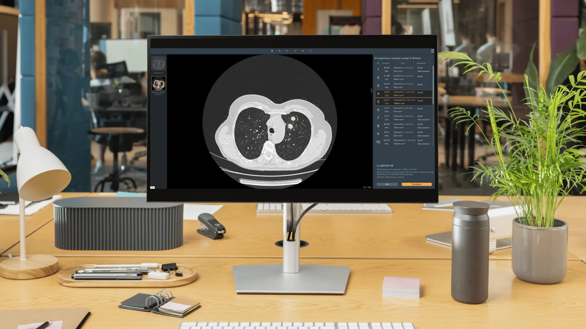

Discover how Rayscape works
integrate Rayscape AI into
your hospital's infrastructure
and improve both your
daily workflow and patients' experience.
