Abstract
Lung nodule detection remains one of the most common and painstaking tasks in radiology. Efforts to aid overworked radiologists are made using artificial intelligence (AI), although computed tomography (CT) makes it a hard computational task in practical scenarios.
This study analyses the translation of a weight-averaging ensemble technique, from natural image classification to small object detection on CT. A dataset of 1050 patients is used to fine-tune models under diverse configurations to compare different types of ensembles.
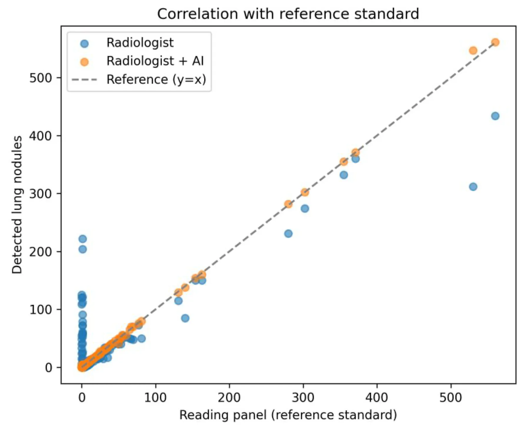
The model soup boosts the FROC score from 0.872 to 0.886, with no computational downsides. Next, two radiologists test their detection performance with and without the ensemble assistance on 20 CT studies. The AI improves the physicians’ mean sensitivity from 91.2 ± 5.1% to 94.2 ± 4.6%, while preserving a non-inferior specificity (P < 0.001). These results further pave the way to translating general computer vision advancements into the medical domain, supporting AI’s place in the physician’s toolbox.
Excited to explore further? Send us an email at [email protected].