

Best AI-Powered Radiology Software in 2025: Part 2
The remarkable synergy between human expertise and AI-powered tools continues to reshape the boundaries of what's achievable in diagnostic imaging. Radiologists feel empowered by these digital companions, seamlessly integrating AI-driven insights into their decision-making process.
In this exploration of the latest AI-powered radiology companies, we uncover a diverse spectrum of advancements that cover everything from bone age evaluation and fracture detection to cardiac analysis and brain imaging. We have traversed the landscapes of lung health, breast cancer screening, and neurological conditions, witnessing the prowess of AI as it aids in the detection, diagnosis, and monitoring of various ailments.
Click here to revisit Part 1: BEST AI RADIOLOGY 2025
As the medical community opens its arms to these AI-driven marvels, we're seeing a shift that's blurring the line between sci-fi and reality. Machines are learning to see the invisible, uncovering patterns once hidden in the shadows. Part 2 of our AI software in Radiology series is a deep dive into the heart of innovation, shining a spotlight on companies that are pushing the boundaries of what's possible in 2025.
Are you excited to catch a glimpse of the radiology of tomorrow? Let's jump in and explore the future together!
Lunit
Lunit Inc. introduces a transformative deep learning-based software, named INSIGHT CXR, which is designed to upgrade radiologists and clinicians in interpreting chest X-rays, specifically in the PA or AP view. This AI-driven solution harnesses the power of automation to detect essential radiologic findings, ranging from atelectasis to pneumothorax. With a notable emphasis on cancer detection, Lunit INSIGHT CXR addresses critical abnormalities, including cardiomegaly, consolidation, and fibrosis. Its impact is structured around three pivotal elements:
- Localization of Suspicious Areas: By accurately pinpointing and highlighting suspicious regions, the software provides a visual guide through color or outlines, instantly drawing attention to potential anomalies.
- Abnormality Scores: Each identified lesion is assigned an abnormality score, quantifying the probability that the detected feature indicates an abnormal condition, adding depth to the analysis.
- Textual Interpretation: A detailed textual interpretation is provided for each finding, offering comprehensive insights into the context and significance of the radiologic discoveries.

Lunit INSIGHT CXR seamlessly integrates into the existing workflow of radiologists, delivering valuable support at crucial stages: Before analysis, the software optimizes worklist orders and swiftly identifies acute findings, streamlining processes and prioritizing cases; During analysis, it serves as an intuitive perception aid, prompting the recognition of abnormalities, displaying results, and offering heatmaps for enhanced interpretation.
Aidoc
With a focus on enhancing both efficiency and accuracy, Aidoc has developed an impressive suite of AI products designed to identify critical abnormalities across the body. These algorithms, meticulously crafted from vast quantities of data, provide indispensable diagnostic aid across a broad spectrum of pathologies. From intracranial hemorrhage to spine fractures, intra-abdominal free gas, rib fracture, and even pulmonary embolism, Aidoc's comprehensive array of algorithms covers diverse areas of medical imaging analysis.
One of Aidoc's groundbreaking contributions to the field is the concept of "Always-on AI". This approach involves the constant analysis of medical imaging data in the background, automatically identifying urgent findings and sparing radiologists from drowning in a sea of irrelevant information. Aidoc's "Always-on AI" serves as a beacon of efficiency, ensuring that critical abnormalities are promptly detected and communicated, enabling timely interventions.

Aidoc enhances healthcare by deploying solutions through its advanced aiOS™ system, revolutionizing radiology with fast integration. Notably, Aidoc's prowess extends to specific solutions tailored to address distinct pathologies. Their Intracranial Hemorrhage (ICH) solution offers triage and notification capabilities, flagging suspected positive findings of this life-threatening condition in non-enhanced head CT images. Similarly, their Pulmonary Embolism (PE) solution is analyzing CTPA images, effectively detecting and communicating cases of pulmonary embolism. Furthermore, Aidoc's brain aneurysm solution provides triage and notification tools for CT images, ensuring the prompt identification and communication of suspected brain aneurysms.
Contextflow
Contextflow, an Austrian startup born from the collaboration between the Medical University of Vienna (MUW) and the Khresmoi European research project, has pioneered a transformative approach in radiology. At its core is the contextflow ADVANCE Chest CT, an innovative 3D image-based search engine meticulously designed for diagnosing lung conditions. With the capacity to identify 19 distinct disease patterns in lung CTs, this technology stands as a visual detective, matching and highlighting relevant disease patterns to guide clinicians in their diagnostic journey.

This intelligent system empowers radiologists by returning meticulously curated reference cases and pertinent medical knowledge, all directly accessible through a smart integration within the PACS. In practice, contextflow ADVANCE Chest CT comes to life during critical decision-making moments, offering interactive support as it reveals abnormalities and results on demand, ultimately revolutionizing the diagnostic process with its user-friendly web-based interface.
Oxipit
Surfacing from Vilnius, Lithuania, Oxipit offers a comprehensive and dynamic platform for medical imaging. At the forefront of their innovative offerings is ChestEye Quality, an automatic Quality Assurance (QA) solution tailored specifically for chest X-rays. This technology enhances the medical team by implementing a dual approach. It meticulously double-reads every chest X-ray, engaging in an intricate analysis of radiologists' reports and juxtaposing them with AI-generated insights. If any potential False Negatives (FN) are detected within the radiologists' diagnoses, ChestEye Quality strategically flags these cases for final review, ensuring a comprehensive evaluation.

With a resolute objective to elevate reporting accuracy and bolster the radiologists' confidence, ChestLink stands as an indispensable safety net, meticulously designed to prevent any possible FNs and enhance the overall quality of diagnoses. Covering a vast spectrum of 75 different pathologies, its autonomous healthy patients' report generation feature adds to its prowess, enabling AI-only diagnosis without the interference of a radiologist. Oxipit's unwavering dedication to accuracy and efficiency positions ChestLink as an essential tool in the arsenal of modern radiology practices.
Qure
Qure, an innovative player in the realm of AI medical solutions based in India, is charting a transformative path across various medical domains, including Lung, Heart, Neuro, and MSK. With a strong focus on enhancing diagnostic capabilities, Qure's AI technologies offer invaluable assistance in critical medical scenarios. In the context of head injuries or strokes, their qER solution takes center stage as a pivotal diagnostic aid, ensuring timely triage and diagnostic support.
By prioritizing the most urgent scans on radiology worklists, qER enables swift reviews, efficiently detecting and localizing critical abnormalities like bleeds, fractures, mass effects, and midline shifts, while quantifying their severity. qXR extends Qure's prowess to chest X-rays, detecting up to 29 common abnormalities and serving as a robust tool for tuberculosis screening. In the pursuit of lung health, qCT-Lung emerges as a vital asset, contributing to lung cancer screening initiatives.

This AI solution not only aids clinicians by swiftly detecting lung nodules and emphysema but also empowers opportunistic screening efforts. Furthermore, Qure's dedication to musculoskeletal health is evident through its qMSK trauma solution, adeptly processing multiple views in under 20 seconds and drawing insights from its extensive training on over 1 million scans.
iCAD
Catering to the needs of radiologists, iCAD introduces ProFound AI™ for 2D Mammography, a proficient solution meticulously designed to analyze each image, proficiently detecting both malignant soft tissue densities and calcifications. Leveraging the power of deep learning and artificial intelligence, ProFound AI for breast tomosynthesis presents a transformative tool that aids radiologists in addressing the intricacies of reading tomosynthesis images. Complementing its lineup, ProFound AI Risk emerges as a valuable clinical decision support tool, offering personalized breast cancer risk estimates over one, two, and three-year spans.
By amalgamating diverse risk factors like age, density, and subtle mammographic features, ProFound AI Risk equips physicians to craft tailored screening plans, a significant stride towards early detection and customized patient care.

Mammography holds paramount importance as a cornerstone of breast cancer screening and early detection efforts. As one of the most effective methods for identifying breast abnormalities, mammography plays a pivotal role in detecting cancer at its nascent stages, when treatment options are most successful and the prognosis is significantly improved.
QUANTIB
Quantib® Prostate is an innovative software that revolutionizes the workflow for detecting abnormalities in MRI prostate scans. This comprehensive solution encompasses a range of key features, including precise prostate gland and zone volume measurements, AI-supported region-of-interest segmentation, calculation of PSA density, and robust support for PI-RADS v.2.1 scoring. Notably, Quantib® Prostate enhances diagnostic accuracy by providing structured reports and aiding radiologists in identifying potential abnormalities through intuitive heatmaps and perception assistance during analysis. Additionally, it offers interactive decision support, presenting results and abnormalities on demand, as well as insightful report suggestions, optimizing the interpretation process.

In a parallel endeavor, Quantib introduces Quantib® ND, an intelligent radiology application that automates brain structure segmentation and quantification. This transformative tool provides valuable insights into conditions like Alzheimer's disease and dementia by assessing atrophy and offering automatic tracking of neurodegeneration over time. Moreover, Quantib® ND facilitates white matter hyperintensity (WMH) detection and quantification, proving particularly beneficial for monitoring patients with multiple sclerosis (MS).
Its ability to distinguish between new and previously identified WMHs, along with color-coded lesion visualization superimposed on anatomical maps, allows for comprehensive analysis. By offering brain structure segmentation, volume quantification, multi-time-point assessments, and precise white matter hyperintensity quantification, Quantib exemplifies innovation in AI-driven radiology solutions.
Rayscape
Rayscape is a complete digital assistant designed to empower doctors with powerful Artificial Intelligence tools, enabling better decision-making and improved patient care. Focused on the health of the lungs, this comprehensive suite of products seamlessly integrates into medical workflows, allowing radiologists to analyze chest X-rays and lung CTs faster and more efficiently.
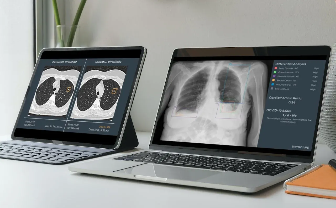
Rayscape CXR, an integral software component, revolutionizes patient triage by automatically distinguishing between normal X-rays and those with anomalies. With the ability to detect over 148 pathologies, it empowers doctors with the following:
- Additional X-ray visualizations for unparalleled accuracy
- Localization of the lesions on the scan
- Calculation of the cardio-thoracic index
- Patient prioritization based on identified pathologies
Rayscape can be installed into the pre-existing infrastructure and flows without disruptions, with fully customizable options for both On-Premise or Cloud integrations.

The stage is set for a new era, one where AI-driven marvels seamlessly collaborate with radiologists, unraveling mysteries hidden within medical images and revolutionizing the way we approach healthcare. The future beckons, brimming with promises of novel perspectives on complex medical conditions. So, as we bid adieu to this captivating exploration, let us stride forward together, embarking on an exciting adventure toward the radiology of tomorrow.
Welcome to the future.
If you want to learn more about Rayscape's custom AI solutions, please contact us here.



Discover how Rayscape works
integrate Rayscape AI into
your hospital's infrastructure
and improve both your
daily workflow and patients' experience.
