

Detecting Early Lung Cancer with AI on Routine X-rays
Lung cancer rarely arrives loudly. It develops in silence, growing behind ribs and over years—quietly outpacing symptoms, eluding early suspicion. And while CT is the recommended tool for screening, many patients don’t make it to that step. They start, and sometimes end, with an X-ray.
It might be a routine check. A pre-op. A cough that doesn’t resolve. And in that image—low resolution, low contrast, imperfectly positioned—might be the first visible sign of something that doesn’t wait.
Radiologists know the odds. They know that X-rays miss early nodules. But they also know that in real-life practice, not everyone gets ideal workup. Not everyone gets the follow-up CT. In these spaces, what matters is not whether the tool is perfect, but whether it’s used perfectly.
Next, you’ll see cases where Rayscape’s AI supported radiologists in finding the subtle, silent beginnings of pathology on chest X-rays—cases that might otherwise have gone unnoticed, or worse, unflagged until too late.
What is Rayscape?
Rayscape is a radiology AI company focused on augmenting radiologists and clinical teams in their daily decision-making. Our technology assists in the interpretation of chest X-rays and lung CT scans, using large-scale medical image training to detect, measure, and monitor key findings.
Designed by radiologists, for radiologists, Rayscape is built with real-world workflow in mind—delivering support where it matters most: during on-call shifts, under pressure, and in the screening or monitoring of oncology patients—environments where speed and accuracy are critical.
Built to support, not replace- Rayscape assists the radiologist in seeing more, seeing earlier, and seeing clearly.
The reality of lung screening
While guidelines point to low-dose CT as the gold standard, many health systems still rely on chest X-rays as a first-line investigation—especially in large population screening, pre-operative workups, or post-pandemic triage.
And in this setting:
- Many scans are read quickly, with low suspicion
- Patient follow-up is inconsistent or delayed
- Early signs are easily masked by anatomy or image quality
- Nodules may be small, subtle, or embedded in expected contours
- Fatigue and volume can impact even the most experienced radiologist’s performance
Here’s how Rayscape makes a difference
- Identifies small nodules and lung lesions that may be missed in low-suspicion reads
- Highlights abnormalities without overriding the radiologist’s judgment
- Ensure consistency in image analysis, regardless of time of day or workload
- Offer structured overlays and measurements, without disrupting the existing workflow
Rayscape doesn’t turn an X-ray into a CT. But it does turn a second’s glance into a more structured review. It helps ensure that if something is there, it has a better chance of being seen—before it becomes something more.
Proven clinical value
In the following, you’ll see clinical cases from screening programs and routine check-ups—real scenarios where Rayscape helped radiologists identify nodules and subtle lesions hidden behind structures on chest X-ray.
Each case is anonymized and presented with AI overlays as they appeared in practice. These are not hypothetical scenarios—they are taken from daily workflows, under real clinical pressure.
#SCREENING CASE 1: What was obvious wasn’t all
Patient: Male, 66 years old
Setting: Screening Program – Routine Chest X-ray
Clinical Presentation: Mild chronic cough, no prior imaging on file
Imaging: Chest X-ray
Clinical Context
Sometimes, screening doesn’t begin with subtlety. Sometimes, the abnormal is obvious. The surprise is what sits beneath it.
He was 66. Still working. Still biking on weekends. The referral read “chronic cough – rule out infection.” No weight loss. No dyspnea. Just a routine scan late in the afternoon—standard protocol for primary care peace of mind.
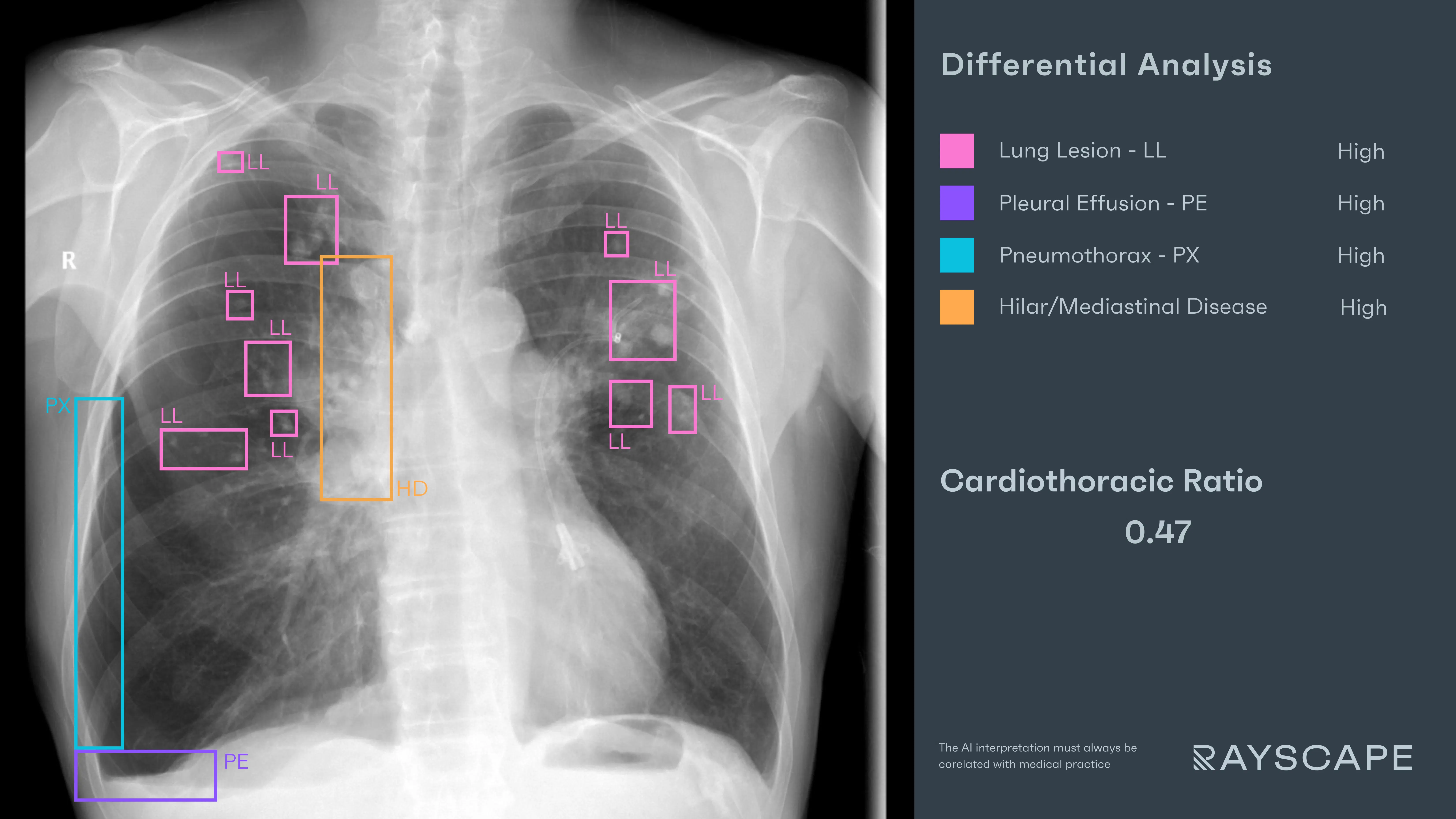
And yet, the image was far from routine. Even at first glance, the right hemithorax was distorted: too much air where there shouldn’t be, too much fluid pressing upward from below. It demanded immediate attention.
But radiologists know this scenario too well: when the pathology is loud—a pneumothorax, an effusion—the eye naturally locks in. The louder the finding, the easier it is for smaller signs to drift beneath awareness. Nodules, especially, disappear under this kind of urgency—not because they’re invisible, but because they’re quiet.
AI Interaction
Rayscape processed the scan and surfaced what the radiologist already suspected:
- "Pneumothorax" – A clean pleural line arcing across the right chest, with a hollow absence of vascularity beyond it.
- "Pleural Effusion" – Layered fluid in the right lower lung, smooth and small in volume.
These findings alone explained the symptoms. But Rayscape continued, placing more overlays that changed the tone of the review:
- "Hilar/Mediastinal Disease" – A soft tissue mass was outlined at the right hilum, suggestive of enlarged lymph nodes or a primary tumor.
- "Lung Lesions" – Small, rounded opacities began to appear across both lungs—some obvious, others partially obscured by the ribs and spine.
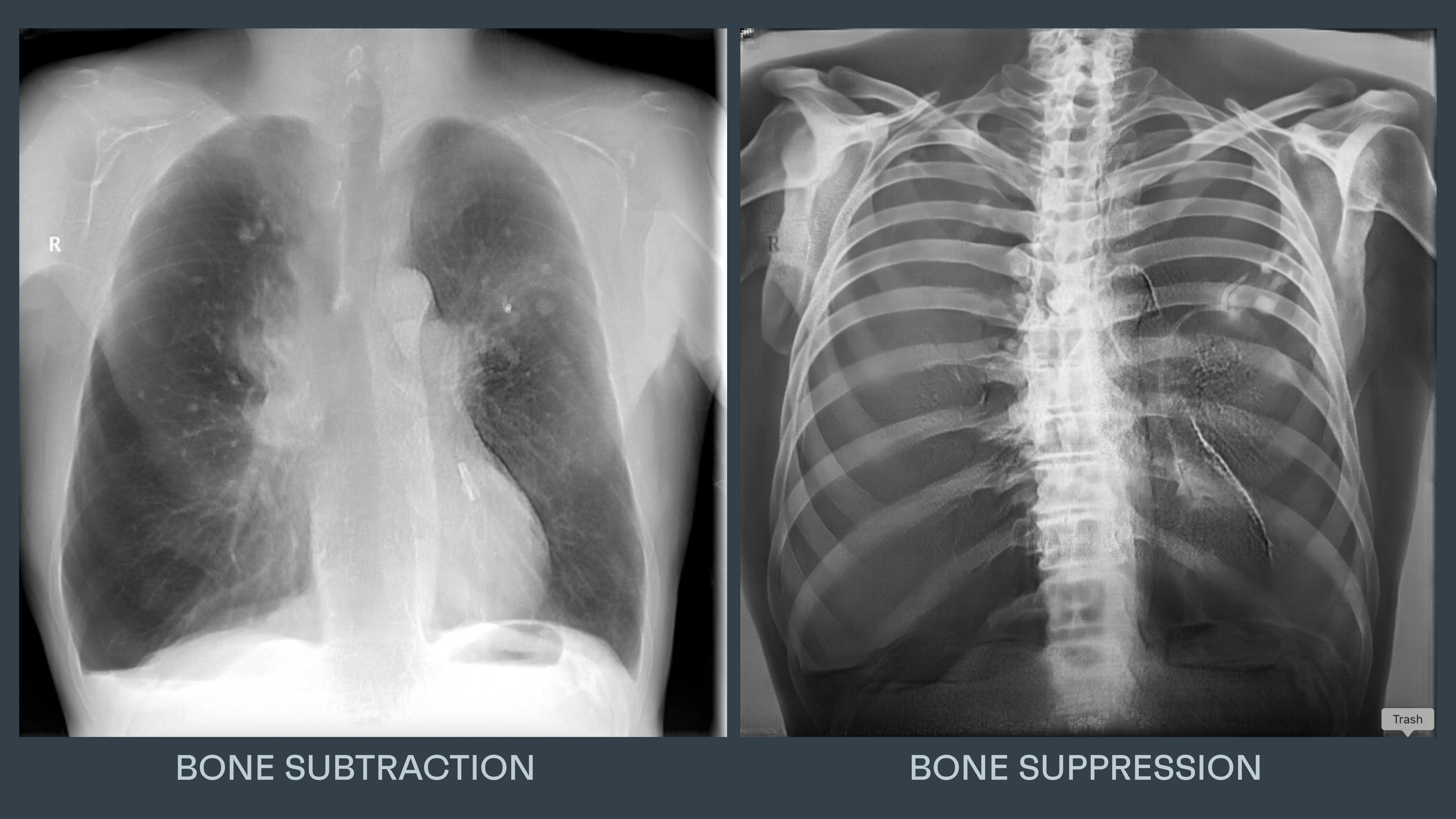
- Bone Suppression + Subtraction Views – When applied, the rib cage faded and the field cleared. Nodules previously hidden became visible: one behind the 4th rib, another near the 5th. Still small, but no longer invisible.
It wasn’t about catching what the radiologist missed. It was about creating room to see more, even when the image was already speaking loudly.
Clinical Response
The tone of the report shifted. “Right pneumothorax and small pleural effusion. Additional findings concerning for underlying malignancy: right hilar fullness and multiple bilateral pulmonary nodules, including several obscured by bone structures and revealed on bone-suppressed views. Recommend urgent CT for further evaluation.”
Outcome
CT confirmed a right hilar mass, mediastinal lymphadenopathy, and scattered bilateral lung nodules consistent with metastatic lung cancer. The pneumothorax and effusion had been only the surface. The underlying story—silent, progressive, and now visible—changed everything.
He had come in for a cough. The scan gave him a diagnosis. But it was the second layer of seeing that gave him a chance.
#SCREENING CASE 2: The rib that hid the nodule
Patient: Male, 61 years old
Setting: Screening Program – Routine Chest X-ray
Clinical Presentation: Asymptomatic, annual check-up
Imaging: Chest X-ray
Clinical Context
The request was standard: “Routine CXR – annual exam.” No complaints. No smoking history noted. No follow-up flagged. The kind of scan that slides through the PACS unnoticed—quiet, unprioritized, surrounded by a dozen just like it.
This is the hidden paradox of screening: the less urgent the case, the more vital the catch. The more normal the context, the easier it is to miss what matters most. There’s no blood pressure to chase. No fever to explain. Just bone, air, and the hope that nothing is hiding.
And yet—something was.
AI Interaction
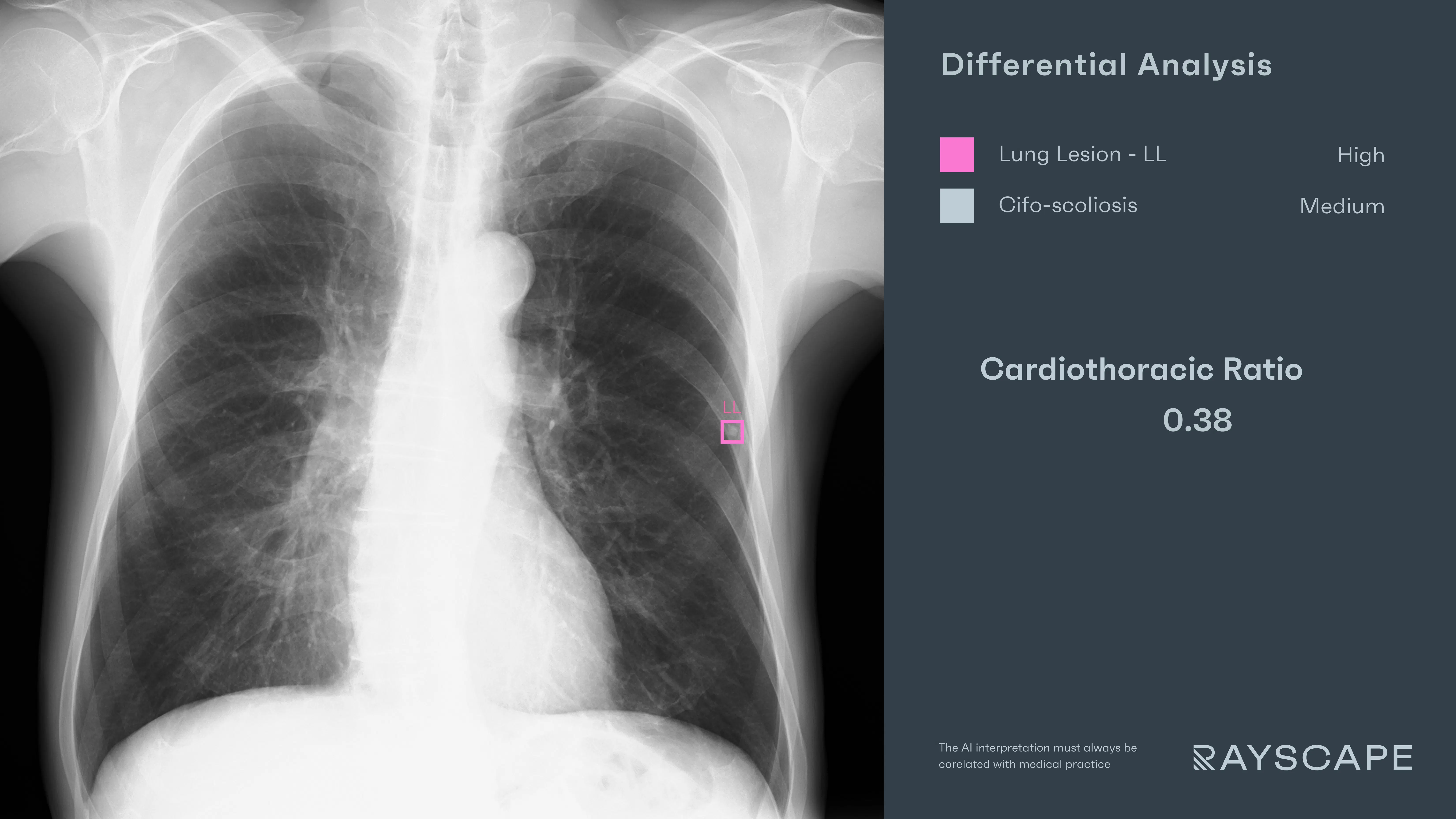
Rayscape processed the image in its usual rhythm, scanning the lungs for asymmetries and contours that broke from expectation. Then it placed a single, subtle overlay:
- "Lung Lesion" – A small, round nodule identified over the posterior arc of the 6th and 7th ribs on the left, partially obscured by bone. It was well-defined but faint—sitting right in the line where rib intersects parenchyma, blending into the anatomical noise.
Without the overlay, the eye might have passed over it entirely. A shadow on a shadow. But the AI paused on it, quietly offering a frame—a question, not an answer.
The radiologist leaned in, adjusted the contrast, and confirmed the contour: a solitary pulmonary nodule, subtle and perfectly placed to avoid suspicion. Had it not been highlighted, it might’ve been filed under “normal.”
Outcome
The report noted: “Small, well-defined left pulmonary nodule projected over rib shadow. Recommend lateral view or CT for further evaluation.”
A follow-up low-dose CT confirmed a 6 mm solid nodule in the posterior segment of the left lower lobe. It was entered into the nodule follow-up protocol, with scheduled imaging over the next two years.
The patient remained asymptomatic. But the path had shifted—because someone, or something, had looked twice.
#SCREENING CASE 3: Faint, but not forgettable
Patient: Male, 50 years old
Setting: Screening Program – Routine Chest X-ray
Clinical Presentation: No symptoms, former smoker, annual exam
Imaging: Chest X-ray
Clinical Context
The difficulty in screening isn’t just volume. It’s repetition. The same silhouettes, the same angles, the same normal lungs—until they’re not.
He was a 50-year-old former smoker. No cough. No weight loss. Just routine surveillance—something his daughter insisted on, since he hadn’t seen a doctor in three years. The referral was clean. The expectation: another scan to file under normal.
The lungs looked clear at first. The film was well-exposed, technically adequate. Nothing alarming. But screening is built on suspicion—not symptoms.
AI Interaction
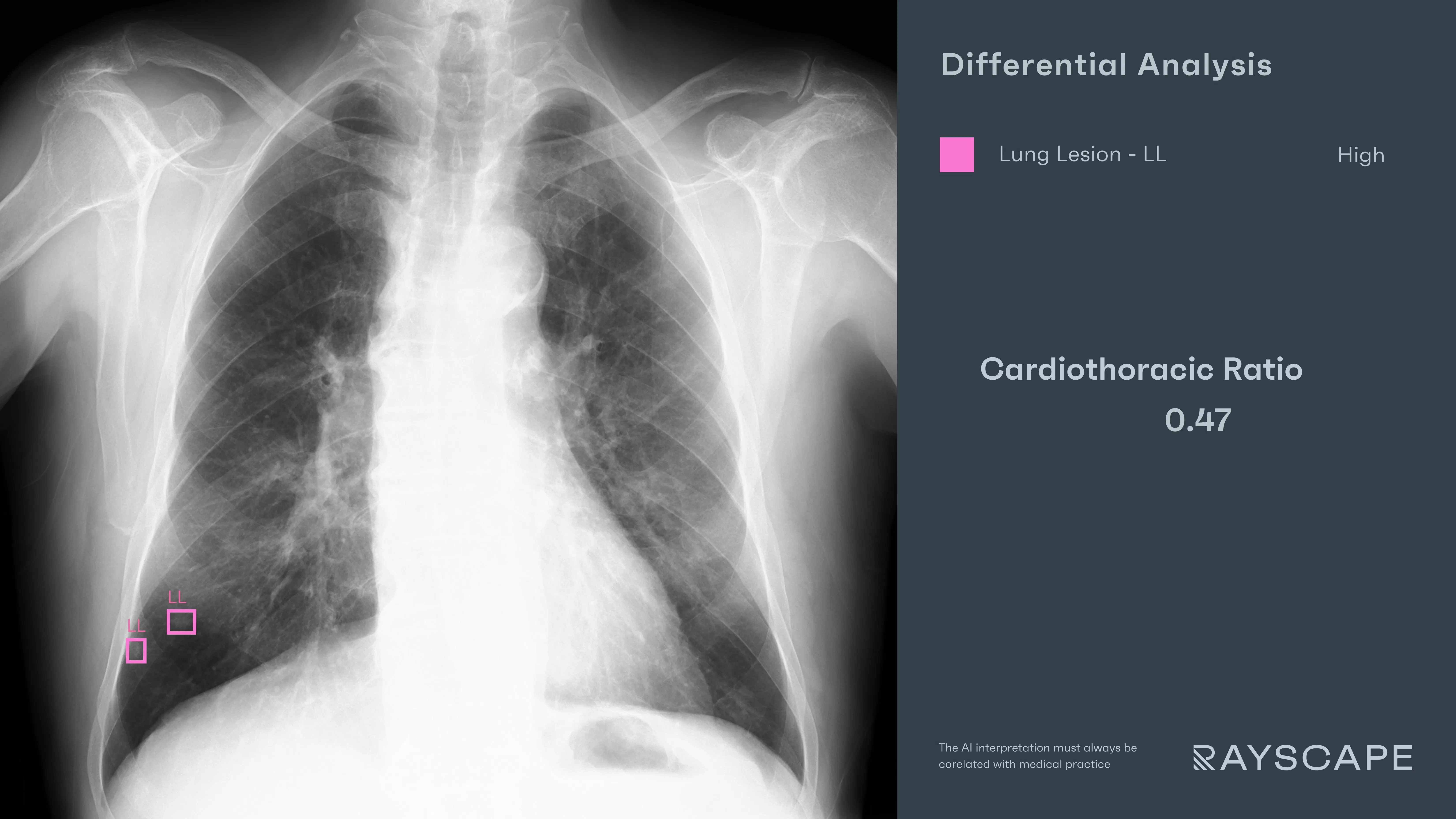
Rayscape’s AI scanned the image in the background, as always. At first glance, nothing stood out. But then two overlays appeared, hovering just above the right hemidiaphragm:
- "Lung Lesion" – One annotation marked a faint, round opacity—subtle enough to vanish in a breath.
- "Lung Lesion" – A second nodule, slightly smaller, adjacent to the right costophrenic angle, blurred by overlapping ribs.
The findings were barely perceptible. Easily dismissed as artifact. But the structured overlay shifted the frame. The radiologist narrowed the window, leaned in, adjusted contrast—and there they were. Still faint. Still easy to doubt. But undeniably there.
These weren’t calcified. They weren’t old scars. And they didn’t belong.
Outcome
The report noted: “Two subtle nodular opacities in the right lower lung field. Recommend low-dose CT for further characterization.”
The CT was performed within a week. It confirmed two solid pulmonary nodules measuring 6 mm and 8 mm, respectively, with spiculated margins and high-risk features. The case was referred to a multidisciplinary oncology team for follow-up.
Discussions
Lung cancer screening doesn’t feel like emergency medicine. There are no beeping monitors or code carts outside the reading room door. Just a queue of quiet patients, and a stack of images that almost all look normal.
And that’s the challenge. In screening, the greatest risk isn’t noise—it’s silence. The nodules are faint. The lungs are clear. The findings don’t shout. They suggest.
For the radiologist, it’s not about missing something obvious. It’s about staying sharp when everything looks nearly normal, hour after hour, film after film. Because sometimes, one faint contour in one lower lobe is the only warning a patient will ever get.
Rayscape doesn’t change the image. It doesn’t tell the radiologist what to think. It offers a nudge—a second look, a tighter frame, a structured prompt to pause before dismissing what looks too small to matter.
In screening, speed is important. But vigilance is everything. And that’s where AI earns its place: not in reading louder, but in helping radiologists see quieter, more clearly, and with more confidence—especially when nothing else seems wrong.
Built to fit your workflow
For IT teams and PACS administrators, implementation is often a key concern. Rayscape is designed to fit into your existing systems:
- Compatibility with major PACS platforms and cloud environments
- Vendor agnostic: trained to work on every DICOM image, no matter the vendor of the machine or the protocol
- Flexible deployment: on-premise, hybrid, or cloud-based
- Various outputs: SC, GSPS, SR
- Fast onboarding and secure data management protocols (HIPAA / GDPR compliant)
- Available on all major marketplaces (Deepc, Blackford, Carpl) or through direct sale
- We work closely with your radiology and IT departments to ensure seamless rollout with minimal disruption.
Let’s Talk
Your team makes life-saving decisions every day. Rayscape is here to support you in those critical moments—bringing clarity, speed, and confidence to emergency imaging.
👉 Contact us and learn how we can empower your radiology team—when it matters most.

Discover how Rayscape works
integrate Rayscape AI into
your hospital's infrastructure
and improve both your
daily workflow and patients' experience.
