

AI for Chest X-ray Interpretation in the Emergency Room
The rising volume of imaging studies in emergency care is creating a new type of challenge: one where demand outpaces attention. While imaging technology has evolved rapidly, the availability of radiologists—and the time they can dedicate per case—remains limited.
This imbalance contributes to an increasing risk of human error, delayed diagnosis, and missed findings, particularly in time-sensitive environments like the Emergency Room.
We invite you to explore real-world clinical scenarios where Rayscape’s AI-powered software appears not as a decision-maker, but as a perceptive observer—illuminating subtle findings, reinforcing attention, and lending a steady hand where the workload strains precision.
What is Rayscape?
Rayscape is a radiology AI company focused on augmenting radiologists and clinical teams in their daily decision-making. Our technology assists in the interpretation of chest X-rays and lung CT scans, using large-scale medical image training to detect, measure, and monitor key findings.
Designed by radiologists, for radiologists, Rayscape is built with real-world workflow in mind—delivering support where it matters most: during on-call shifts, under pressure, and in the screening or monitoring of oncology patients—environments where speed and accuracy are critical.
Built to support, not replace- Rayscape assists the radiologist in seeing more, seeing earlier, and seeing clearly.
Saving time, saving lives - the role of AI in the Emergency Room
In the Emergency Department, every second counts. Radiologists must assess a high number of studies under strict time constraints, often with incomplete clinical data and limited imaging quality.
Clinicians rely on fast reporting for life-threatening decisions. And the reality is that:
- Portable X-rays often produce suboptimal images
- Overlapping structures can mask subtle findings (pneumothorax, effusions, nodules)
- Fatigue and volume can impact even the most experienced radiologist’s performance
Here’s how Rayscape makes a difference
- Support radiologists with instant AI-generated highlights that draw attention to overlooked or evolving findings
- Help prioritize urgent cases based on potential risk
- Ensure consistency in image analysis, regardless of time of day or workload
- Offer structured overlays and measurements, without disrupting the existing workflow
With Rayscape, radiologists can focus on what they do best—making life-saving decisions—while we help ensure nothing critical gets missed.
Proven clinical value
In the following, you’ll find real clinical cases from emergency departments across different institutions—each one showcasing how AI has supported radiologists in detecting, confirming, and communicating urgent findings such as:
- Pneumothorax
- Fractures
- Lung infiltrates
- Pleural effusions
...and more.
Each case is anonymized and presented with AI overlays as they appeared in practice. These are not hypothetical scenarios—they are taken from daily workflows, under real clinical pressure.
#ER CASE 1: Subtle lines, serious signals
Patient: Female, 36 years old
Setting: Emergency Room
Clinical presentation: Sudden onset dyspnea, mild tachypnea, slight hypoxia, normotensive
Imaging: Chest X-ray
Clinical Context
A 36-year-old female patient is brought to the emergency department by ambulance following a sudden onset of dyspnea while at work. She is alert, oriented, and speaking in short sentences. Her respiratory rate is elevated, and her oxygen saturation is dipping slightly below normal on room air. The physical exam is inconclusive: no crackles, no obvious asymmetry, diminished breath sounds—but not strikingly so.
The attending physician, considering a wide differential—from pulmonary embolism to viral bronchitis—orders a portable chest X-ray.
Behind the scenes, the radiology team is contending with the familiar pressures of a late-shift ER. Cases arrive in waves. Trauma, stroke alerts, incidental findings, endless portables. Fatigue seeps into the reading room like background radiation—never quite seen, always present.
There is a rhythm to reviewing images quickly but thoroughly. But in this rhythm, there are blind spots. Not due to incompetence or neglect, but due to the cognitive overload that builds slowly across every shift. A line too fine, a fluid level too subtle, a pattern too familiar to raise alarms. These are not "misses"—they are the margins where human attention can fray under pressure.
AI Interaction
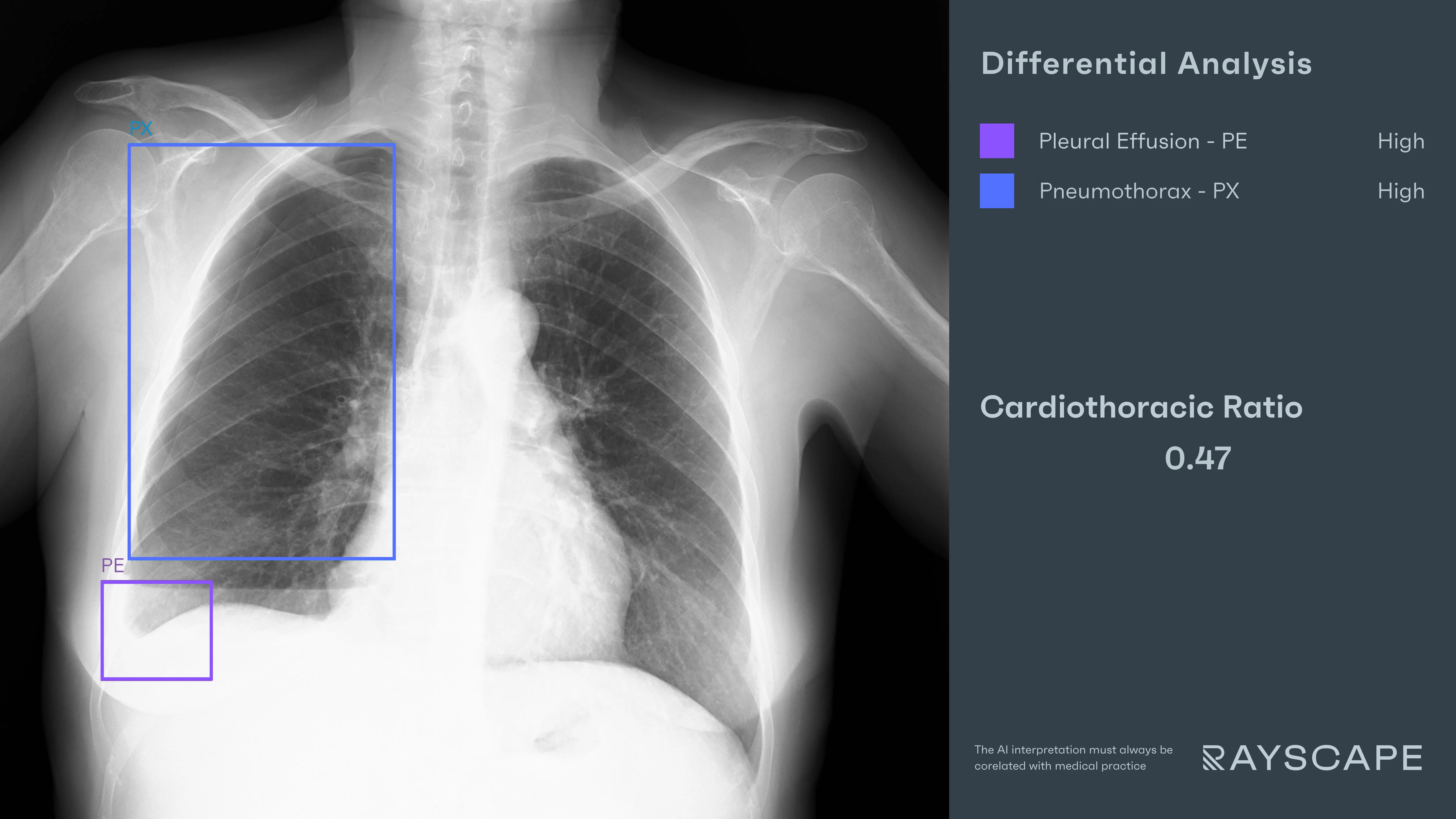
As the chest X-ray loads into the PACS viewer, the Rayscape AI assistant runs its analysis in parallel. Within seconds, its interface overlays two distinct bounding boxes:
- "Pneumothorax" – A precise box drawn along the right hemithorax, highlighting the fine pleural line where lung markings abruptly disappear. A thin, easily overlooked contour.
- "Pleural Effusion" – A second bounding box is placed over the right costophrenic angle, pointing out the blunting of the contour and a subtle visible meniscus, suggesting moderate fluid accumulation.
The AI does not diagnose. It does not shout "hydropneumothorax" in capital letters. But it quietly places both findings in the radiologist’s line of sight—structured, consistent, and visually defined.
The radiologist, experienced and sharp despite the hour, had initially considered the possibility of effusion. But with the AI overlay prompting a second glance at the opposing lung, she pieces together the full picture.
The combination of findings—the air above, the fluid below—forms a classical hydropneumothorax, a duality that could easily be filed under “mild” in the absence of supportive tools.
Outcome
The radiologist updates the report in real time, adding the new finding. The ER physician is contacted directly. Bedside ultrasound confirms both air and fluid in the pleural cavity. A chest tube is placed under sterile conditions, and the patient stabilizes within the hour.
#ER CASE 2: The Space Between the Heart and the Lungs
Patient: Male, 72 years old
Setting: Emergency Room
Clinical Presentation: Generalized fatigue, orthopnea, peripheral edema, mildly altered mental status
Imaging: Portable Chest X-ray
Clinical Context
He arrived from the care facility just past midnight, curled under a thin blanket and a vague referral note: “fatigue, O₂ sat low, consider fluid overload.” His breathing was shallow, his blood pressure high, and his gaze slow to follow the room. The nurse noted JVD and soft bilateral crackles. This is the kind of case that feels familiar until it isn’t.
The physician ordered a portable chest X-ray. The image arrived as expected: slightly rotated, a little underpenetrated. And yet, it carried enough to raise a question, if someone knew where to look.
AI Interaction
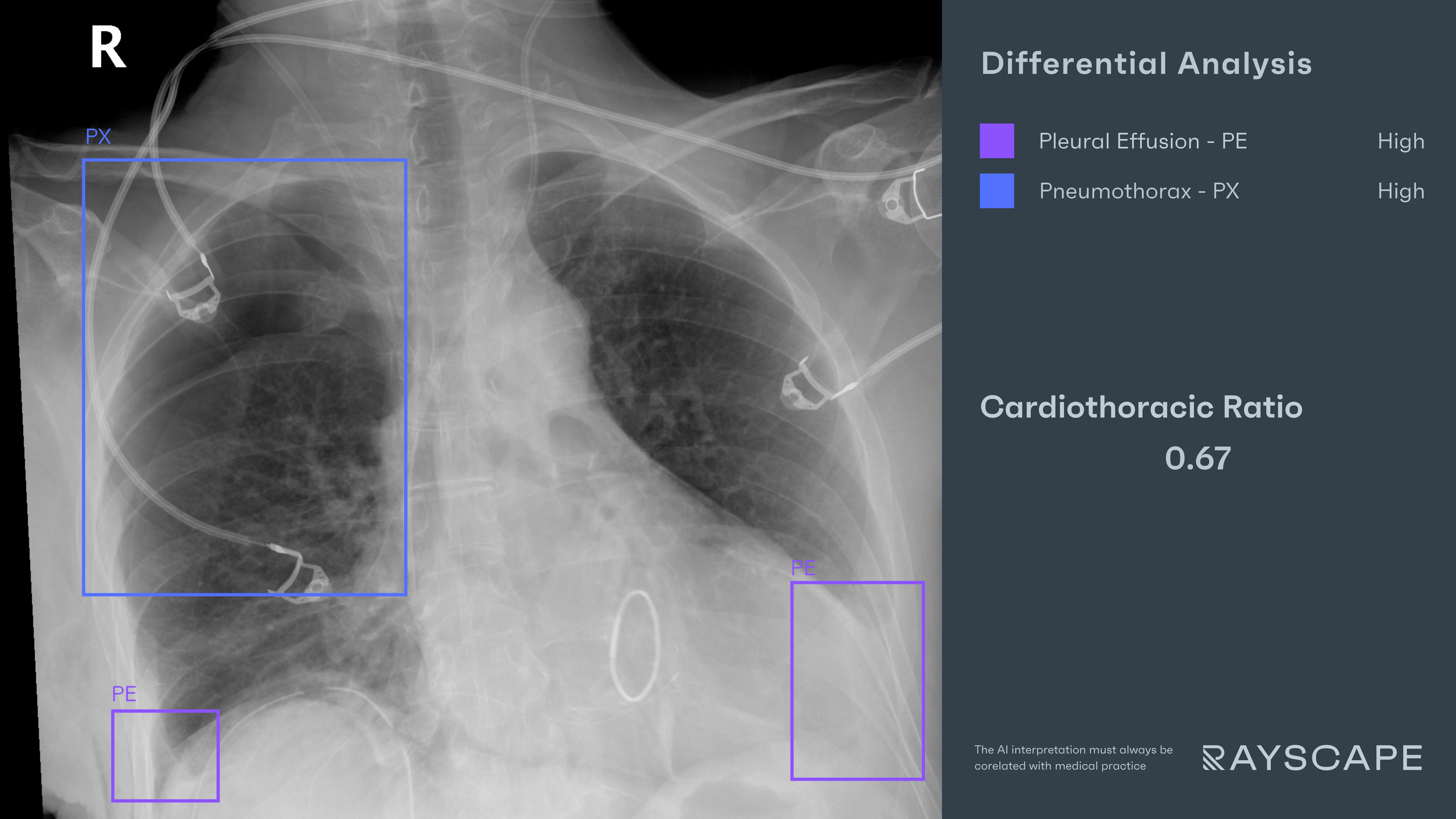
As the image loaded, Rayscape’s AI moved ahead—quietly reading in parallel, not to conclude, but to frame.
- "Right Pneumothorax" – A bounding box traced the upper right hemithorax. The pleural line, sharp but narrow, ran parallel to the chest wall.
- "Pleural Effusion" – Bilateral boxes hovered near the costophrenic angles. The classic meniscus sign bowed gently upward on both sides, more generously on the left.
- "Cardiothoracic Ratio: 0.67" – A third overlay emerged across the cardiac shadow: a subtle annotation, quiet and numerical, signaling an enlarged silhouette.
These were not loud alerts. They were fragments. But together, they gave shape to a story: cardiomegaly, bilateral effusions, and a right-sided pneumothorax—three elements rarely aligned by accident.
The radiologist paused, adjusted windowing, and rewound her thinking. What looked like a simple case of fluid overload now suggested acute decompensated heart failure, complicated by hydropneumothorax on the right.
Outcome
The report was updated promptly. The emergency physician called for a point-of-care ultrasound and a cardiology consult. A right-sided drain was placed, relieving the air. Diuresis followed. The patient stabilized, no longer drifting under the weight of hidden fluid and air.
#ER CASE 3: Collision at the Ribcage
Patient: Male, 40 years old
Setting: Emergency Room – Trauma Bay
Clinical Presentation: Blunt chest trauma from a high-speed motor vehicle collision
Imaging: Chest X-ray, followed by Chest CT
Clinical Context
The impact was front-end, 90 km/h. He was restrained, conscious, and in pain. His right chest rose unevenly, marked with subcutaneous bruising and tenderness across the lateral ribs. Vitals wavered—hypotensive, tachypneic, O₂ at 90% on mask.
The trauma team moved quickly. Chest X-ray first. CT pending. The image came through fast, raw and imperfect, like most in the trauma bay.
AI Interaction
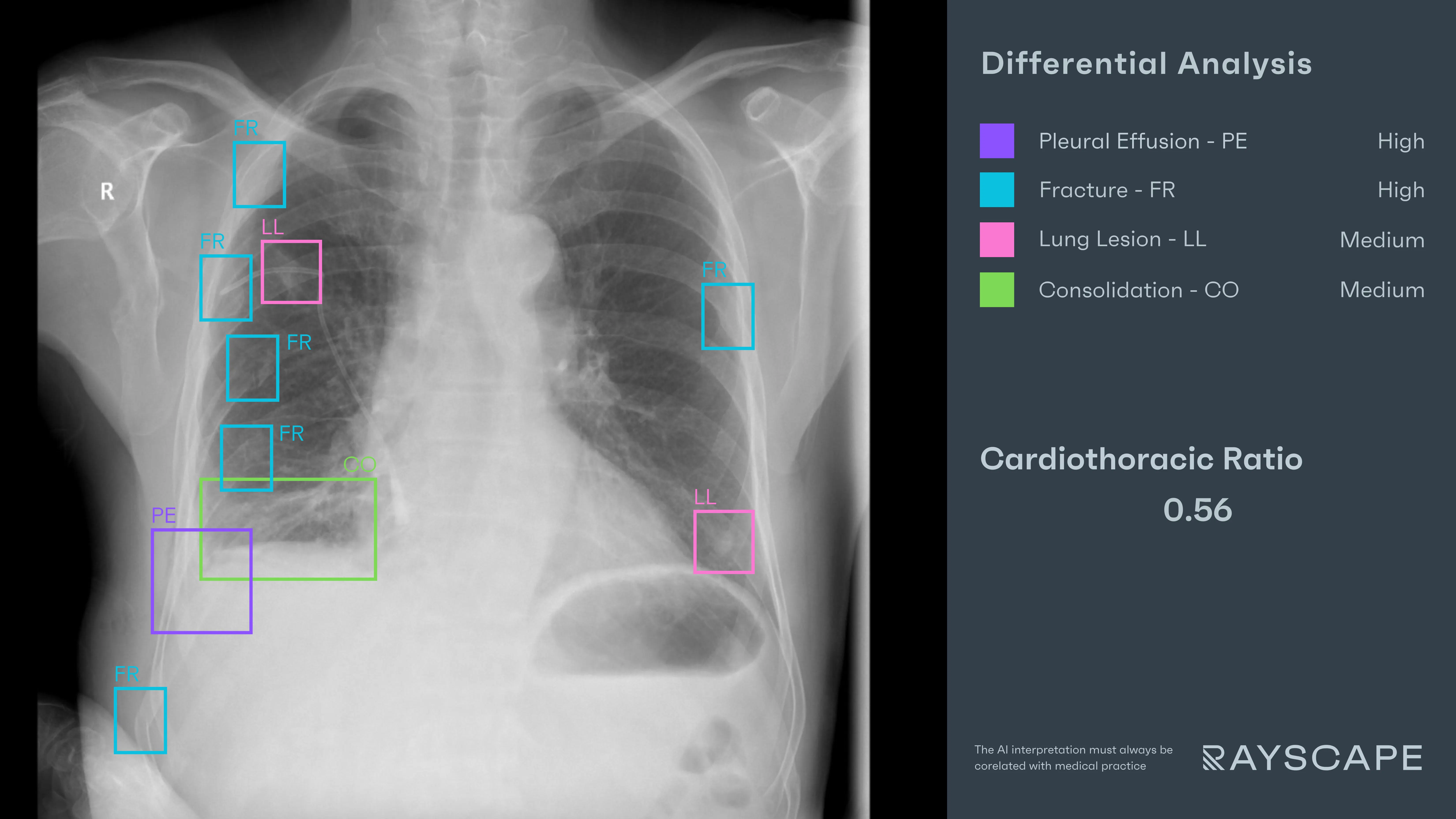
Rayscape stepped in—not to diagnose, but to steady the frame, to sort the noise from the signal.
- "Multiple Rib Fractures" – Bounding boxes outlined multiple ribs on the right and also on the left, displaced and sharply angulated. One had pierced the soft shadow of the intercostal space.
- "Pleural Effusion" – A soft opacity pooled low, where the diaphragm faded and the spine rose—a probable hemothorax, in this setting.
- "Lung Opacity" and “Consolidation” – A final box wrapped the lower right lobe, irregular and dense, textured like wet paper: pulmonary contusion or early consolidation.
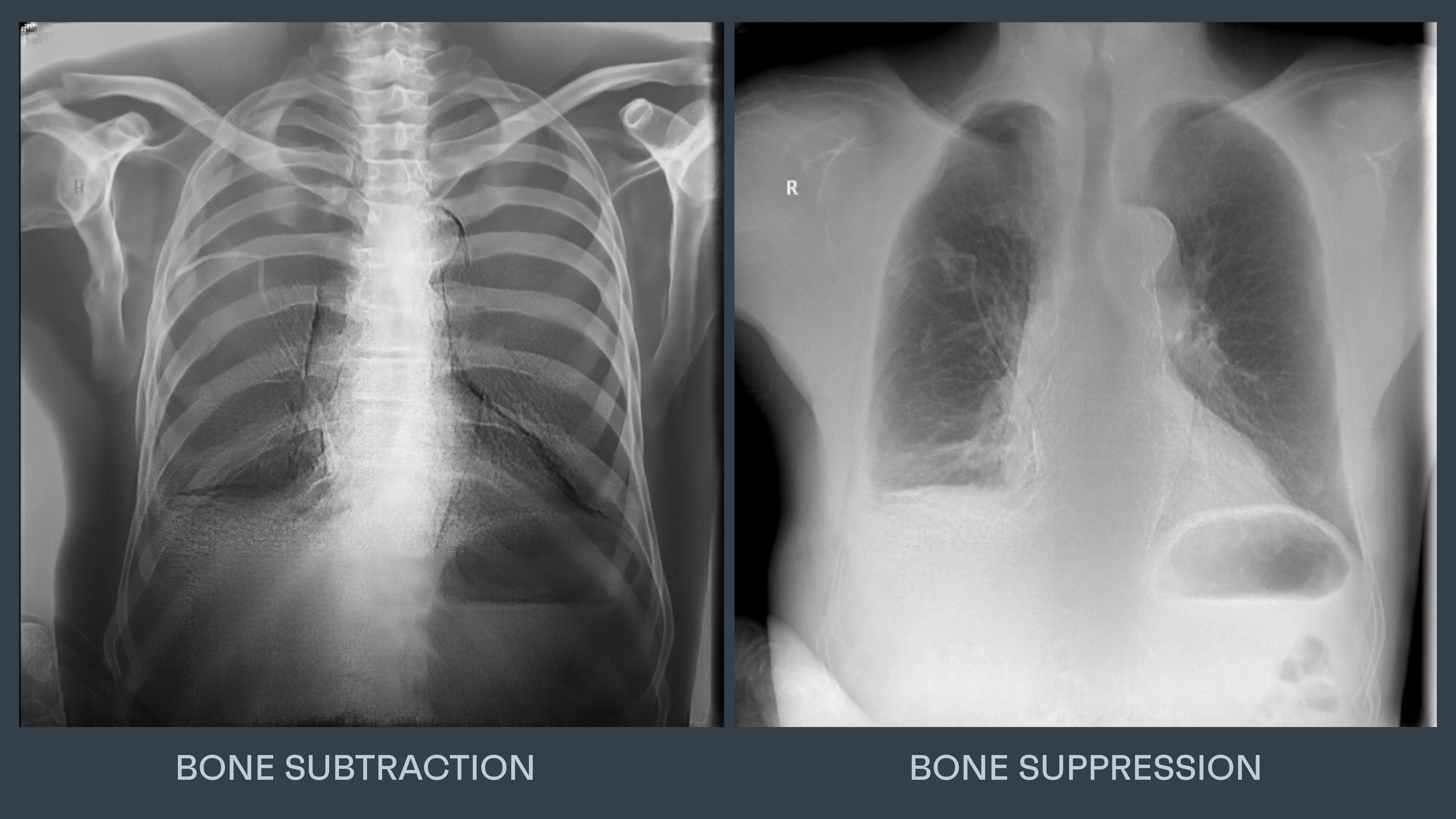
The initial image was complex. So, the AI applied bone suppression, stripping back the ribs to let the lung speak for itself. Then came bone subtraction, peeling away lung artifacts and confirming the multiple fractures.
It wasn’t just about detection. It was about visual translation—about giving the radiologist the language to describe trauma before the scan arrived.
Outcome
CT confirmed the full picture: multiple rib fractures, hemothorax, and lung contusions. A chest tube was placed. The patient was stabilized and admitted to the trauma ward for monitoring and pain management.
Discussions
The practitioners are not tireless, infallible, or immune to distraction; behind every report is a human reader, juggling uncertainty, probability, and the relentless tick of the clock.
In these cases, the pathology wasn’t buried. It wasn’t hidden. It simply existed in that grey space where fatigue, volume, and subtle presentation blur the signal.
The AI didn’t “catch” what the radiologist missed—it offered clarity before the fog could form. It positioned the findings within the frame, allowing the mind to move faster, the pattern to resolve more sharply.
And this is the quiet revolution: not replacement, but reinforcement. Not autonomy, but augmentation. Rayscape acted not as a second opinion, but as a second set of steady hands in a moment where one pair may not be enough.
The future of ER imaging doesn’t look like machines reading scans alone. It looks like this: a collaborative frame, where human intuition meets algorithmic precision. Where a bounding box becomes not a conclusion, but a question, well-posed, in just the right place.
Built to fit your workflow
For IT teams and PACS administrators, implementation is often a key concern. Rayscape is designed to fit into your existing systems:
- Compatibility with major PACS platforms and cloud environments
- Vendor agnostic: trained to work on every DICOM image, no matter the vendor of the machine or the protocol
- Flexible deployment: on-premise, hybrid, or cloud-based
- Various outputs: SC, GSPS, SR
- Fast onboarding and secure data management protocols (HIPAA / GDPR compliant)
- Available on all major marketplaces (Deepc, Blackford, Carpl) or through direct sale
- We work closely with your radiology and IT departments to ensure seamless rollout with minimal disruption.
Let’s Talk
Your team makes life-saving decisions every day. Rayscape is here to support you in those critical moments—bringing clarity, speed, and confidence to emergency imaging.
👉 Contact us. Let’s talk about how we can empower your radiology team—when it matters most.

Discover how Rayscape works
integrate Rayscape AI into
your hospital's infrastructure
and improve both your
daily workflow and patients' experience.
