

Artificial intelligence for lung nodules
A 55-year-old male patient presented to the emergency department with breathing difficulties. The patient's medical history was significant for a 20-year history of smoking.
The initial chest X-ray revealed:
- Enlarged lower left bow in the heart,
- Parietal calcifications in the aortic button,
- Pulmonary hilums increased in volume
The Rayscape CXR identified and notified the radiologists about a relatively well-defined homogenous opacity in the right paratracheal area, which required further imaging investigations. Additionally, there was a hypertransparent strip without pulmonary pattern, located apically and basally on the left lung, with a thickness of approximately 1.7 cm.
The patient was provisionally diagnosed with a tumoral formation and pneumothorax.
Further investigations with CT scan revealed a left pneumothorax with a thickness of up to 2.2 cm. Rayscape Lung CT identified a heterogeneous formation with mixed densities (fluids, parafluids, and solids) measuring approximately 5.6/5.9 cm at the upper right mediastinal level, that surrounded the superior vena cava and stenosed it.
The patient also had pulmonary parenchymal infiltrate at the level of the right upper lobe, and multiple formations with similar imaging appearance observed bilaterally prevascular and hilar up to 2.7cm.

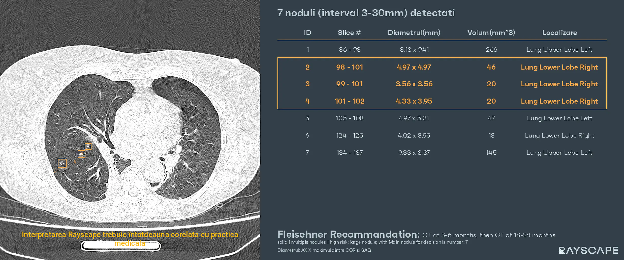
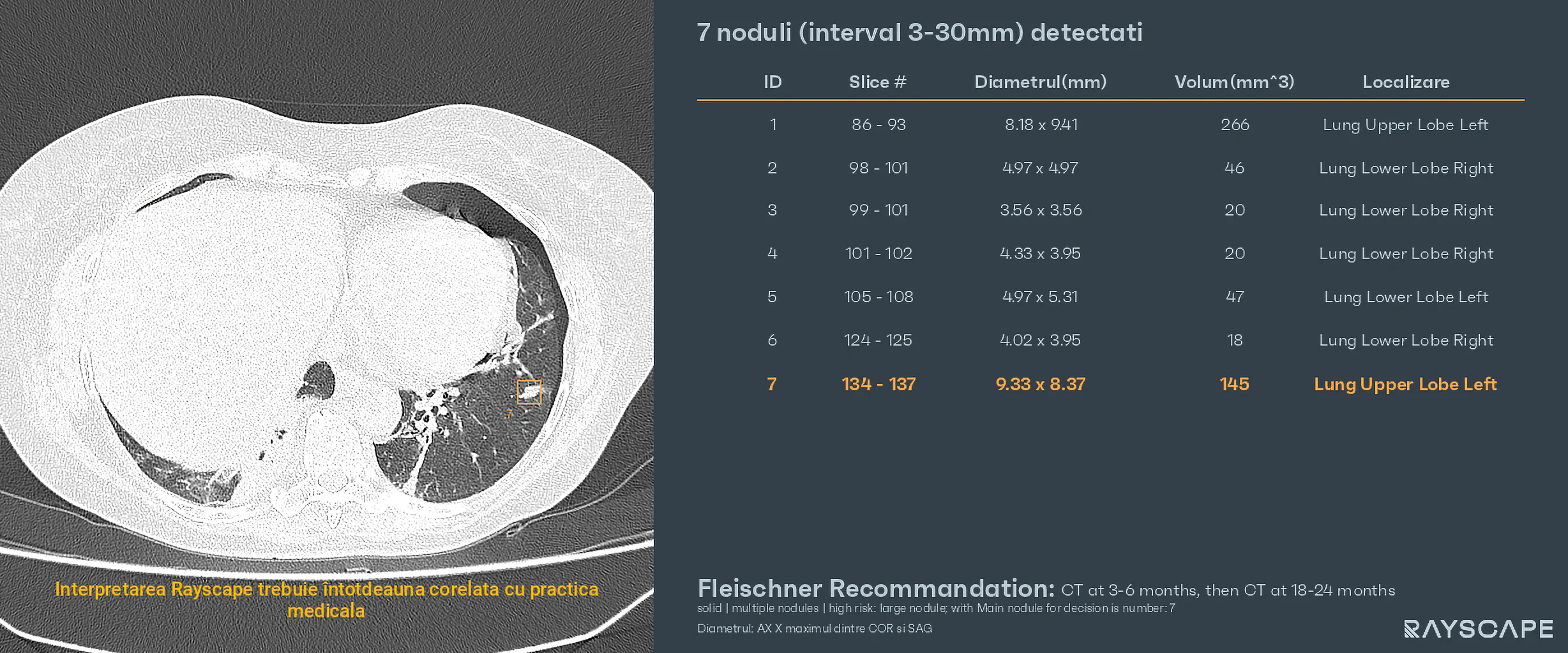
The patient was treated for his emergency condition and advised to undergo further testing to evaluate the lung nodules. However, the patient failed to return for the additional testing.
Two months later, the patient returned to the emergency department with chest pain and dyspnea. A lung CT scan was performed, and the Rayscape comparative analysis tool was utilized.
Compared to the previous investigation, the following findings were observed:
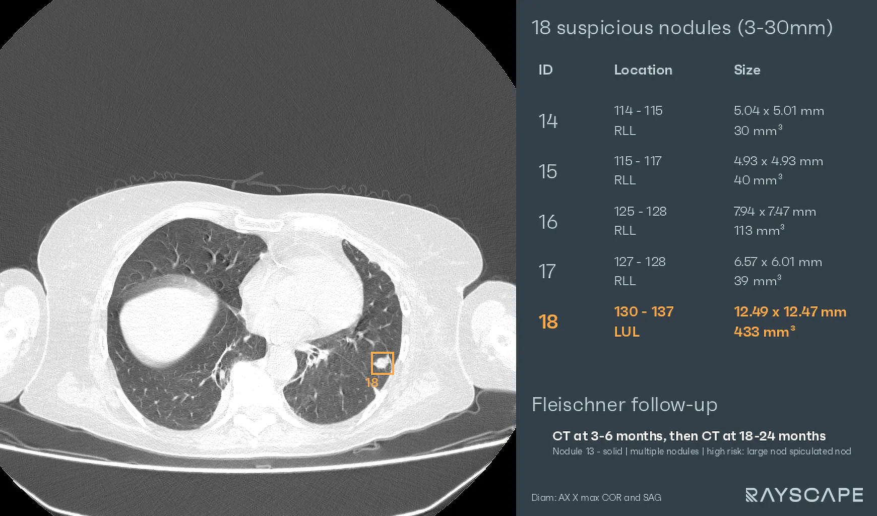
- Mild increase in the upper right lobe tissue formation, predominantly secondary to the presence of air-filled lesions inside, most likely due to erosion of a bronchus and communication with it.
- The formation extends slightly towards the right retroclavicular soft tissues and invades them.
- Mild increase in the size of the paraaortic lymph nodes, as well as the presence of a voluminous secondary adrenal determination bilaterally.
- Mild increase in the size of the previously visualized nodules, as well as the appearance of several pulmonary nodules in both lung fields, the largest being an apical upper left lobe of 8/8mm.
- The excavated basal lesion on the left currently has a tissue aspect.
- The lower right lobe condensation area has a relatively stable appearance.
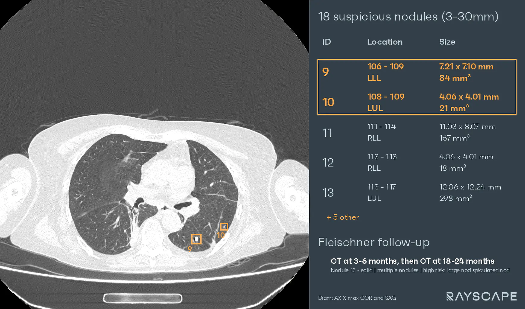
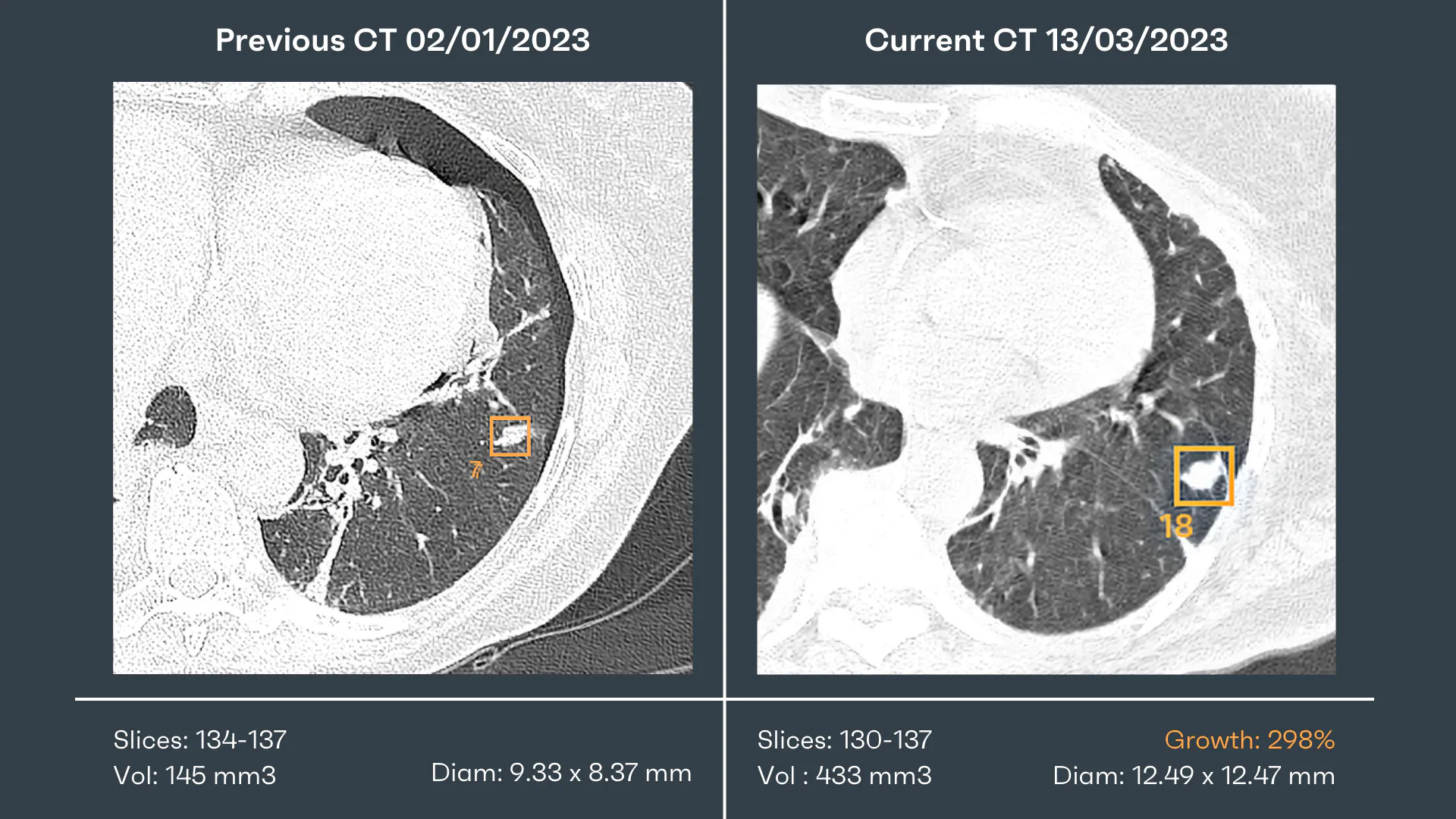
Based on the imaging findings, the patient was diagnosed with lung cancer, and appropriate treatment was initiated.
In this case, the Rayscape comparative analysis tool was crucial in detecting the growing nodules, which were suspected to be malignant lesions, even when the patient failed to return for additional testing. The early detection of lung cancer enabled doctors to provide timely and accurate diagnosis and treatment, ultimately improving the patient's outcomes.

Discover how Rayscape works
integrate Rayscape AI into
your hospital's infrastructure
and improve both your
daily workflow and patients' experience.
