

Pneumothorax in a trauma patient
Patient data
A male patient is admitted to the emergency room with multiple injuries on the upper extremity of the body. His symptoms include sudden chest pain and shortness of breath, with tachypnea and tachycardia. Radiography is used as the first imaging examination method.
Case discussion
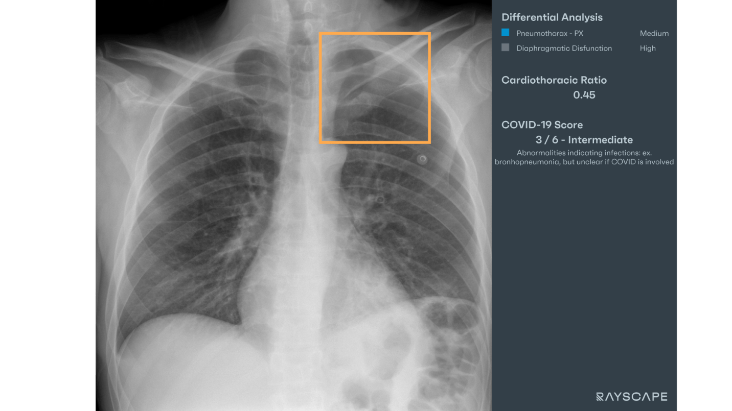
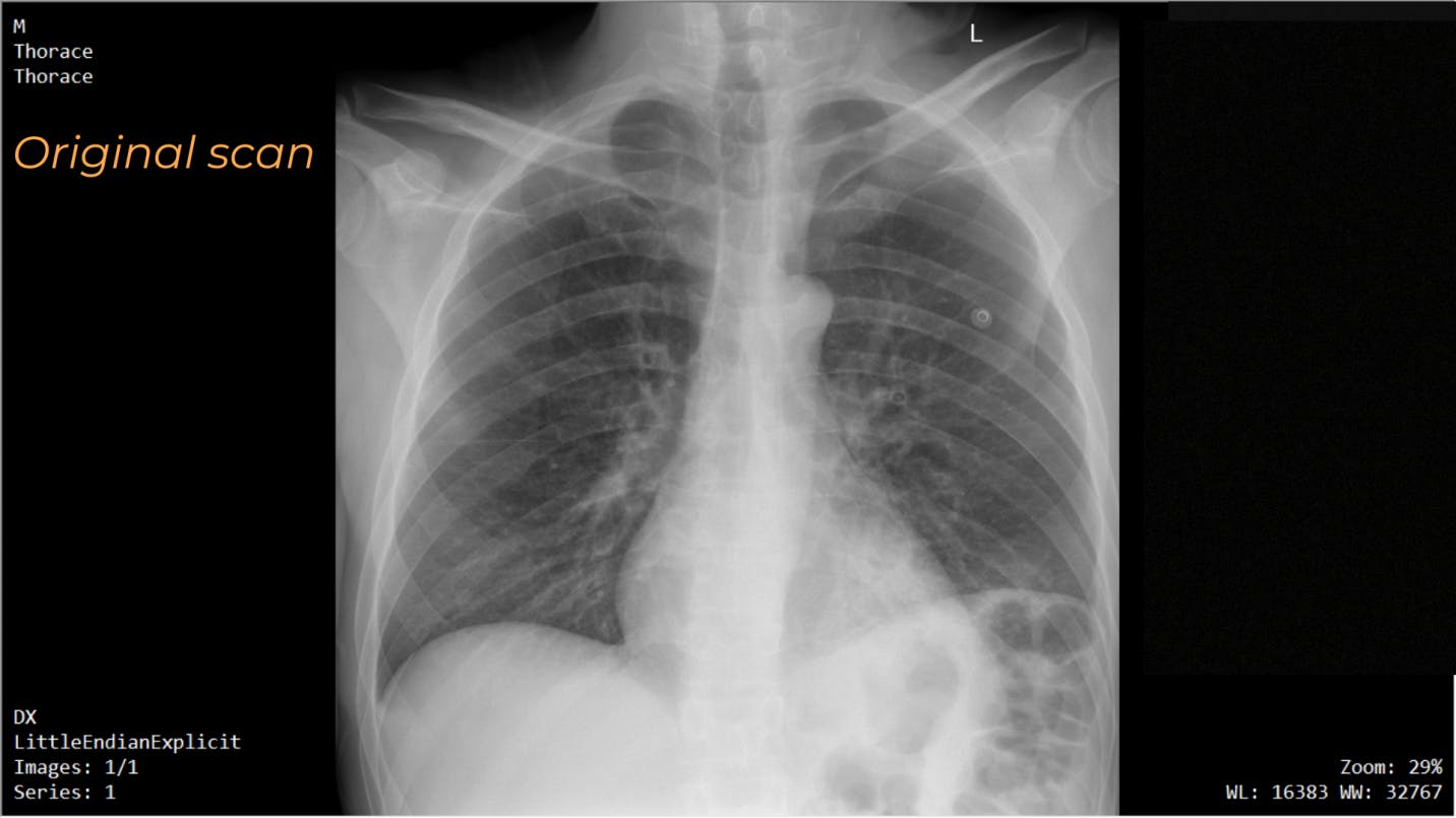
At first glance, the chest X-ray seems normal, but after viewing the Rayscape analysis, our attention is drawn to a possible pneumothorax.
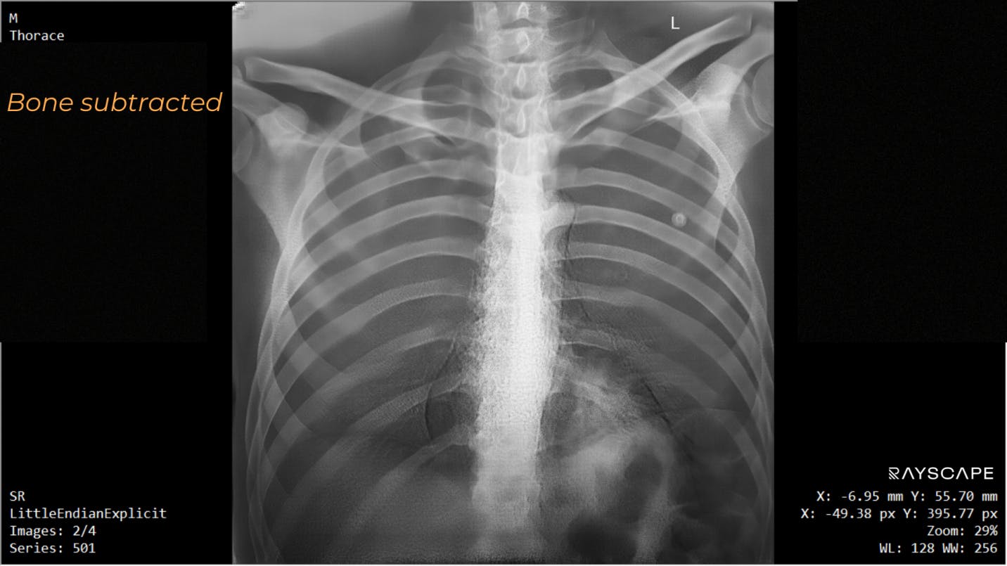
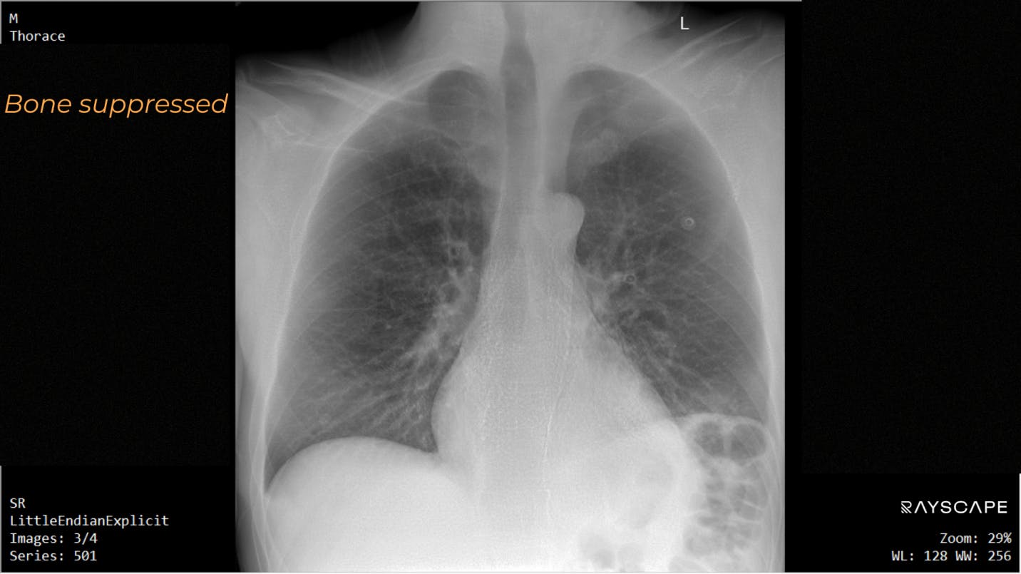
Careful analysis of the radiograph revealed left pneumothorax(left apical radiolucent area with a thickness of approx. 24mm delimited inferiorly by the visceral pleura). A slightly elevated left hemidiaphragm is also observed.
More info
Pneumothorax, sometimes abbreviated to PTX, (plural: pneumothoraxes) refers to the abnormal presence of gas in the pleural space. When this collection of gas is constantly enlarging with resulting compression of mediastinal structures, it can be life-threatening and is known as a tension pneumothorax (if no tension is present it is a simple pneumothorax). An occult pneumothorax refers to a missed one on initial imaging, usually on a supine/semierect chest radiograph.
Possible treatment:
- Observation
- Supplemental oxygen
- Needle aspiration
- Chest tube drainage:
- Chemical pleurodesis
- Surgery: Video-assisted thoracoscopic surgery (VATS)
References
1. "What Are the Signs and Symptoms of Pleurisy and Other Pleural Disorders". www.nhlbi.nih.gov. 21 September 2011. . www.nhlbi.nih.gov. 21 September 2011. Archived from the original on 8 October 2016. Retrieved 31 October 2016.
2. Bintcliffe O, Maskell N (May 2014). "Spontaneous pneumothorax". BMJ. 348: g2928.
3. Morjaria JB, Lakshminarayana UB, Liu-Shiu-Cheong P, Kastelik JA (November 2014). "Pneumothorax: a tale of pain or spontaneity". Therapeutic Advances in Chronic Disease. 5 (6): 269–273. "Pneumothorax: a tale of pain or spontaneity". Therapeutic Advances in Chronic Disease. 5 (6): 269–273. . Therapeutic Advances in Chronic Disease. 5 (6):
4. Weinberger S, Cockrill B, Mandel J (2019). Principles of Pulmonary Medicine (7th ed.). Elsevier. pp. 215–216. ISBN 9780323523714 9780323523714.
Discover how Rayscape works
integrate Rayscape AI into
your hospital's infrastructure
and improve both your
daily workflow and patients' experience.
